

 النبات
 الحيوان
 الأحياء المجهرية
 علم الأمراض
 التقانة الإحيائية
 التقنية الحيوية المكروبية
 التقنية الحياتية النانوية
 علم الأجنة
 الأحياء الجزيئي
 علم وظائف الأعضاء
 الغدد
الغدد
 المضادات الحيوية
|
Read More
Date: 28-7-2016
Date: 28-7-2016
Date: 8-1-2017
|
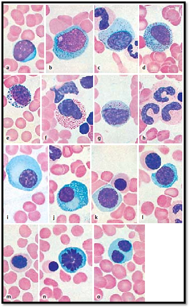
Blood-Sternal Biopsy Smear
a) Myeloblast. The eccentric nucleus leaves only a small seam of strongly basophilic cytoplasm without granules. Note the web-like chromatin structure and the nucleoli.
b) Promyelocyte. The basophilic cytoplasm shows a fine granulation. A nucleus with nucleolus is present.
c) Promyelocyte. The cytoplasm shows only slight granulation. The nucleus is smaller than the one in figure b. It has a coarse structure (myelocyte?).
d) Myelocyte. The cell has become smaller during maturation. It has an oval, coarsely structured nucleus. The cytoplasm contains specific granules.
e) Basophilic myelocyte with granules (stained dark violet).
f) Eosinophilic myelocyte or metamyelocyte, respectively, the nucleus showing the beginnings of a lobed structure. There are eosinophilic granules.
g) Neutrophilic metamyelocyte. The slightly basophilic cytoplasm shows neutrophilic granules.
h) Neutrophilic granulocytes with banded nuclei and coarsely structured chromatin. The cell bodies show beginning granulation.
i) Plasma cell. The nucleus is small and shows hyperchromatosis of the nuclear membrane. The chromatin bodies have a circular symmetry (wheel-spoke nucleus). The basophilic cell body shows a lighter stained cytocentrum (area close to the nucleus).
j) Reticulum cell with basophilic cell body, cytocentrum, numerous fat vacuoles and small coarsely structure d nucleus.
k) Proerythroblast (pronormoblast), a normoblast above it. Proerythrocytes are the largest cells in erythropoiesis (diameter 14–17 μm). Their nuclei dis-play a finely meshed chromatin structure. The basophilic cytoplasm reacts slightly polychromatic.
l) Proerythroblast (center image), macroblast (top), neutrophilic granulocyte with rod-shaped nucleus (right bottom). Note the oxyphilic cytoplasm of the macroblast.
m) Macroblast with almost homogeneous pyknotic nucleus. The cytoplasm is still slightly polychromatic.
n) Mitosis (equatorial plate) in a proerythroblast. The cytoplasm is basophilic.
o) Mitotic macroblast.
Stain: Pappenheim (May -Grünwald, Giemsa); magnification: × 900

References
Kuehnel, W.(2003). Color Atlas of Cytology, Histology, and Microscopic Anatomy. 4th edition . Institute of Anatomy Universitätzu Luebeck Luebeck, Germany . Thieme Stuttgart · New York .


|
|
|
|
علامات بسيطة في جسدك قد تنذر بمرض "قاتل"
|
|
|
|
|
|
|
أول صور ثلاثية الأبعاد للغدة الزعترية البشرية
|
|
|
|
|
|
|
مدرسة دار العلم.. صرح علميّ متميز في كربلاء لنشر علوم أهل البيت (عليهم السلام)
|
|
|