


 النبات
 الحيوان
 الأحياء المجهرية
 علم الأمراض
 التقانة الإحيائية
 التقنية الحيوية المكروبية
 التقنية الحياتية النانوية
 علم الأجنة
 الأحياء الجزيئي
 علم وظائف الأعضاء
 الغدد
الغدد
 المضادات الحيوية
|
Read More
Date: 17-11-2016
Date:
Date: 15-11-2016
|
Division Pteridophyta
Aside from the arthrophyte line of evolution out of the trimerophytes, probably two more led to the plants that dominate Earth at present: ferns (division Pteridophyta) and seed plants.
Ferns first appeared in the Devonian Period and then diversified greatly (Figs. 1 and 2). The division Pteridophyta is still large, with many modern families, genera, and at least 10,000 species (Table ). Ferns can be found in almost any habitat. Moist,
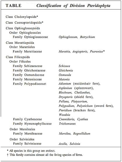
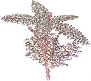
FIGURE 1:This reconstruction of a fern ancestor, Rhacophyton, shows spirally arranged branch systems that had evolved nearly to the point of being called leaves. Note that some are fertile with sporangia and some are sterile; the former evolved into sporophylls, the latter into foliage leaves.
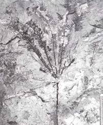
FIGURE 2: A later fern, Phlebopteris, from the Triassic Period. Even though the Triassic occurred from 230 to 190 million years ago, plants had evolved so many modern features that this fossil is not an ancestor to ferns in general, but to a particular family of living ferns, the Matoniaceae. (Courtesy of T. Delevoryas, University of Texas, and R. C. Hope)
shady forests and lakesides are often considered "typical" fern habitats, but species of Woodsia and Cheilanthes occur in dry, hot deserts; Salvinia and Azolla grow floating on water; and Ceratopteris lives submerged below water (Fig. 3). Other genera contain epiphytes (Polypodium) or vines (Lygodium). All ferns are perennial and herbaceous; more is woody, but some do achieve the size of small trees—the tree ferns Angiopteris, Cyathea, Cnemidaria, and others. Although called tree ferns, they never have secondary xylem.
The fern sporophyte consists of a single axis, either a vertical shoot or a horizontal rhizome, that bears both true roots and megaphyllous leaves. The vascular system of the stem is an endarch siphonostele, a derived trait similar to that of division Arthrophyta and the seed plants (Fig. 4). At each node, a leaf trace diverges from the siphonostele, leaving a small segment of the vascular cylinder as just parenchyma; this region is a leaf gap. A vascular cambium has been reported to occur in one fern, Botrychium.
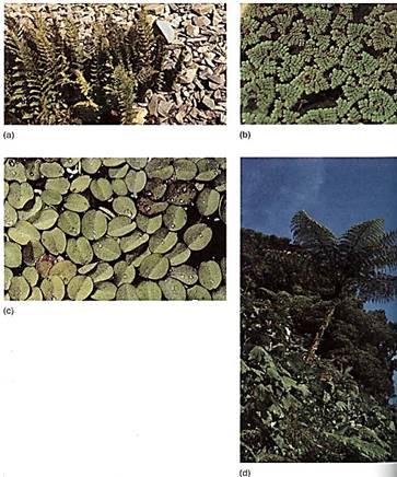
FIGURE 3: (a) Many ferns grow in hot, dry climates with little rainfall. If transplanted to habitats often described as typical for ferns—cool, moist, and shady—these would die within days. Azolla (b) and Salvinia (c) do not look like ferns at first glance, but the structure of their leaves and sporangia shows that they are ferns. (b and c, W. E. Ferguson) (d) Several genera of ferns such as this Cyathea are referred to as tree ferns because they have very long vertical trunks and huge fronds. However, unlike trees, these never have secondary growth and are actually giant herbs. (Patti Murray/Earth Scenes)
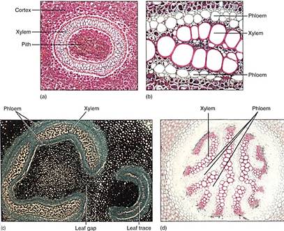
FIGURE 4: Anatomy of fern stems is variable—not only from species to species, but also within a single plant. (a) In this Dennstaedtia and many other ferns, the xylem forms a complete ring in transverse section. (b) Phloem occurs on both the exterior and interior of the xylem in ferns; phloem interior to xylem is extremely rare in seed plants (X 20). (c) In this Adiantum, the vascular tissue makes almost a complete cylinder. A leaf gap occurs where vascular tissue moves to the leaves. Leaf gaps are characteristic of ferns; they do not occur in the microphyll line of evolution (lycophytes). (d) Members of the microphyll line of evolution, such as this Lycopodium annotinum, do not have pith (X 20).
Leaves of ferns may be leathery or delicate, only one cell thick in the filmy ferns, but many layers thick in most. They have an upper layer of palisade parenchyma and a lower region of spongy mesophyll. The leaves are small (Trichomanes) or up to several meters long (tree ferns), but they are almost always compound with a rachis and leaflets (Fig. 5). The staghorn fern (Platycerium) and bird's-nest fern (Asplenium) have simple leaves. Fern leaf primordia have a distinct apical cell, unlike the leaves of seed plants, and as the primordium grows, it curves inward, producing a young leaf that is tightly coiled and commonly known as a fiddlehead (Fig. 6). As the leaf expands, it uncoils and becomes flat. Fern leaves, especially the larger ones, usually contain a considerable amount of vascular tissue. The veins typically branch dichotomously; they are never parallel as in the monocots. Although a type of reticulate venation does occur in a few ferns, it appears more like an imperfect dichotomous venation than the true reticulate venation of dicots.
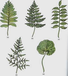
FIGURE 5:Fern leaves (fronds) have a petiole and a lamina, the lamina being almost always pinnately compound. The midrib is a rachis, and the leaflets may also be compound.
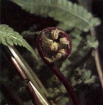
FIGURE 6:Fern leaves have a highly characteristic development: The young leaves are tightly coiled and uncurl as they expand. This is true not only of the rachis, but of the leaflets as well. This circinate vernation does not occur in non-fern plants. (Courtesy of S.-C. Hsiao, University of Texas).
In some species, for example Blechnum spicant and cinnamon fern (Osmunda cinnamomea), certain leaves serve only for photosynthesis and others serve only as sporophylls, but in most ferns, each leaf does both (Fig. 7). On the underside of the leaf are sori (sing.: sorus), clusters of sporangia where meiosis occurs (Fig. 8). Most ferns are homosporous; only two groups of water ferns (Marsileales and Salvineales) are heterosporous.
When fern spores germinate, they grow into small, simple heart-shaped or ribbon-shaped photosynthetic gametophytes with unicellular rhizoids on the lower surface but with no vascular tissue and no epidermis (Fig. 9). Each usually bears both antheridia and archegonia. Antheridia develop early and can be difficult to detect because they occur among the rhizoids; archegonia develop later, close to the gametophyte apex. When the environment is sufficiently moist, antheridia release motile sperm cells that could easily be mistaken lor unicellular green algae except for the absence of plastids. These swim to the archegonia and fertilize the egg.
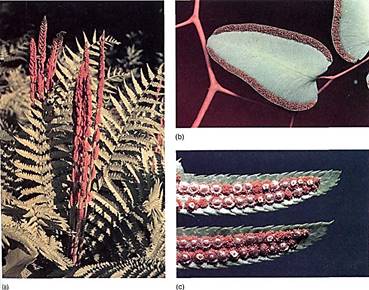
FIGURE 7: (a) This cinnamon fern (Osmunda cinnamomea) contains both foliage leaves that never bear sporangia and highly distinct sporophylls that do bear sporangia. (John N. A. Lott/Biological Photo Service) (b) Most ferns, such as this Pellaea ovata, have only one type of leaf; its broad, thin shape is selectively advantageous for photosynthesis, and it bears sporangia on the underside. (Robert and Linda Mitchell) (c) In almost all ferns such as this sword fern (Polystichum), the sporangia occur in dusters, sori, covered by an indusium. (W. E. Ferguson).
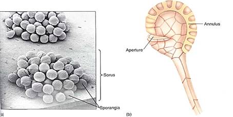
FIGURE 8: (a) A sorus contains many sporangia, each of which consists of a stalk and the actual sporangium body where meiosis occurs. (Biophoto Associates/'Photo Researchers, Inc.) (b) The sporangium opens when specialized cells of the annulus dehydrate, shrink, and suddenly crack open, expanding rapidly and throwing the spores (X 40).
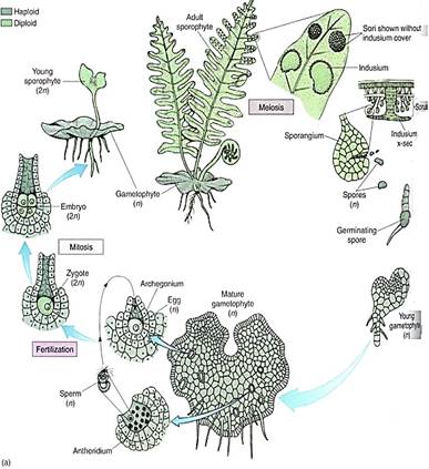
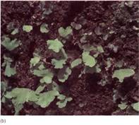
FIGURE 9 (a) Life cycle of a fern. Details are given in the text. (b) Fern gametophytes, several with the first leaves of young sporophytes. (Courtesy of S.-C. Hsiao, University of Texas).
As in Equisetum, the zygote of a fern is retained by the gametophyte that nourishes it. However, the young sporophyte soon produces its first leaves and roots and becomes independent. Its continued growth eventually destroys the small gametophyte. Fern reproduction, with swimming sperms, is still aquatic, but the plants are able to grow in almost any environment as long as there can be at least a temporary high density of gametophytes and a film of dew or rainwater.
Eusporangia and Leptosporangia. Ferns contain two types of sporangia that differ in fundamental aspects of their development. The eusporangium is initiated when several surface cells undergo periclinal divisions, resulting in a small multilayered plate of cells (Fig. 10). The outer cells develop into the sporangium wall, and the inner cells proliferate into sporogenous tissue. This results in a relatively large sporangium with many spores. Anthers and ovules of flowering plants develop this way as well, and the eusporangium is considered to be the basic type in vascular plants.
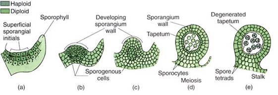
FIGURE 23.40:Development of eusporangia. (a) Surface cells undergo divisions and the inner daughter cells become sporogenous. (b to e) Further divisions result in a large number of spores and a thick sporangium wall.
Leptosporangia are initiated when a single surface cell divides periclinally and forms a small outward protrusion (Fig. 11). This undergoes several more divisions, which result in a small set of sporogenous cells and a thin covering of sterile cells. The leptosporangium produces just a few spores.
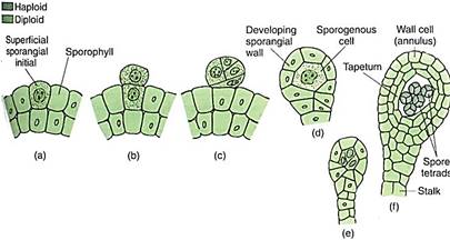
FIGURE 11 :Development of leptosporangia. (a and b) The sporangium is initiated by division in just a single surface cell. (c to f) After a few cell divisions, the sporangium has a stalk, a thin wall, and only a few spores.
Because most plants have eusporangia whereas leptosporangia occur only in some ferns, we believe that having eusporangia is a relictual condition. Leptosporangia must have evolved as a derived condition after the early ferns had come into existence. At present only two orders of ferns, Ophioglossales and Marattiales, still retain eusporangia, whereas all other living ferns are leptosporangiate .
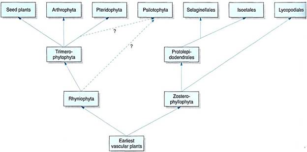
The plants of this chapter—vascular plants without seeds—show great diversity, which at first may seem overwhelming. However, there are only two fundamental lines of evolution out of the rhyniophytes/zosterophyllophytes: the microphyll line (lycophytes) and the megaphyll lines (all the rest except Psilotum; Fig. 12). The second line originated in rhyniophytes, produced the trimerophytes, then split into arthrophytes, ferns, and seed plants.


|
|
|
|
تفوقت في الاختبار على الجميع.. فاكهة "خارقة" في عالم التغذية
|
|
|
|
|
|
|
أمين عام أوبك: النفط الخام والغاز الطبيعي "هبة من الله"
|
|
|
|
|
|
|
قسم شؤون المعارف ينظم دورة عن آليات عمل الفهارس الفنية للموسوعات والكتب لملاكاته
|
|
|