آخر المواضيع المضافة

 علم الكيمياء
الكيمياء التحليلية
الكيمياء الحياتية
الكيمياء العضوية
الكيمياء الفيزيائية
الكيمياء اللاعضوية
مواضيع اخرى في الكيمياء
الكيمياء الصناعية
علم الكيمياء
الكيمياء التحليلية
الكيمياء الحياتية
الكيمياء العضوية
الكيمياء الفيزيائية
الكيمياء اللاعضوية
مواضيع اخرى في الكيمياء
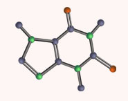
الكيمياء الصناعية | The X–H region (4000–3000 cm–1) distinguishes C–H, N–H, and O–H bonds |


|
|
أقرأ أيضاً
التاريخ: 2-10-2018
التاريخ: 30-3-2017
التاريخ: 30-12-2021
التاريخ: 2023-08-21
|
The reduced masses of the C–H, N–H, and O–H combinations are all about the same. Any difference between the positions of the IR bands of these bonds must then be due to bond strength. In practice, C–H stretches occur at around 3000 cm−1 (although they are of little use in identifying compounds, it’s a rare organic compound that has no C–H bonds), N–H stretches occur at about 3300 cm−1, and O–H stretches higher still at around 3500 cm−1. We can immediately deduce that the O–H bond is stronger than N–H, which is stronger than C–H. IR is a good way to measure such bond strengths.

The form of the absorption bands resulting from X–H IR stretches are very different in these four compounds. Have a look at the shaded portions of the following spectra:


The IR peak of an NH group looks different (spectrum 1) from that of an NH2 group (spectrum 2). A bond gives an independent vibration only if both bond strength and reduced mass are different from those of neighbouring bonds. In the case of an isolated N–H group, this is likely to be true and we usually get a sharp peak at about 3300 cm−1, whether the NH group is part of a simple amine (R2NH) or an amide (RCONHR). The NH2 group is also independent of the rest of the molecule, but the two NH bonds inside the NH2 group have identical force constants and reduced masses, and so vibrate as a single unit. Two equally strong bands appear: one for the two N–H bonds vibrating in phase (symmetric) and one for the two N–H bonds vibrating in opposition (antisymmetric). The antisymmetric vibration requires more energy and is at slightly higher frequency.
The O–H bands occur at higher frequency, sometimes as a sharp absorption at about 3600 cm−1. More often, as in spectra 3 and 4, you will see a broad absorption at anywhere from 3500 to 2900 cm−1. This is because OH groups form strong hydrogen bonds that vary in length and strength. A sharp absorption at 3600 cm−1 indicates a non-hydrogen-bonded OH group; the lower the absorption frequency the stronger the H bond. Alcohols form hydrogen bonds between the hydroxyl oxygen of one molecule and the hydroxyl hydrogen of another. These bonds are variable in length (although they are usually rather longer than normal covalent O–H bonds) and they slightly weaken the true covalent O–H bonds by varying amounts. When a bond varies in length and strength it will have a range of stretching frequencies distributed about a mean value. Alcohols, including the phenol shown in spectrum 3, typically give a rounded absorption at about 3300 cm−1 (contrast the sharp shape of the N–H stretch in the same region you see in the spectra above). Carboxylic acids (RCO2H) form hydrogen-bonded dimers with two strong H bonds between the carbonyl oxygen atom of one molecule and the acidic hydrogen of the other.
These also vary consider ably in length and strength, and usually give the very broad V-shaped absorbance you see in the benzoic acid spectrum 4.

The spectra of paracetamol and BHT (which you met on pp. 58–59) illustrate the effect of hydrogen bonding on peak shape. Paracetamol has a typical sharp peak at 3330 cm−1 for the N–H stretch and then a rounded absorption for the hydrogen-bonded O–H stretch from 3300 down to 3000 cm−1 in the gap between the N–H and C–H stretches. By contrast, BHT has a sharp absorption at 3600 cm−1 as the two large t-butyl groups prevent the typical hydrogen bond from forming.


You may be confused the fi rst time you see the IR spectrum of a terminal alkyne, R–C≡C–H, because you will see a strongish sharp peak at around 3300 cm−1 that looks just like an N–H stretch—the spectrum below (of methyl propynoate, also known as methyl propiolate) illustrates this.
The displacement of this peak from the usual C–H stretch at about 3000 cm−1 cannot be due to a change in the reduced mass and must be due to a marked increase in bond strength. The alkyne C–H bond is shorter and stronger than alkane C–H bonds.

● Typical peak shapes and frequencies for X–H bonds in the region 4000–3000 cm−1.


|
|
|
|
علاج انسداد شرايين الرقبة.. دراسة تنسف "المعتقد الشائع"
|
|
|
|
|
|
|
دراسة علمية تحذر من علاقات حب "اصطناعية" ؟!
|
|
|
|
|
|
|
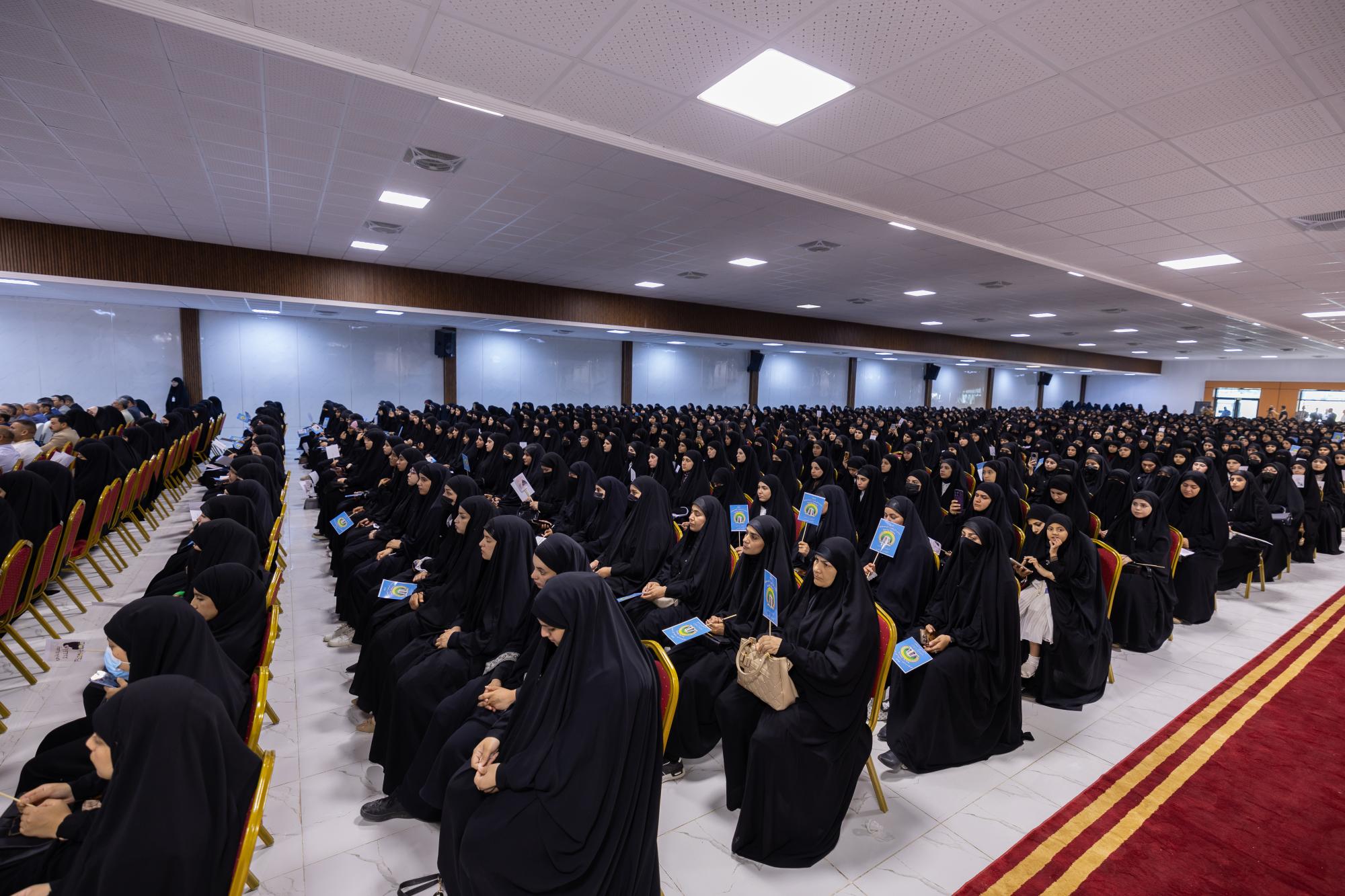
طالبات جامعة واسط: بارتدائنا العباءة نبيّن بأنّ إرث السيدة الزهراء (عليها السلام) ممتد للأجيال
|
|
|