

 النبات
 الحيوان
 الأحياء المجهرية
 علم الأمراض
 التقانة الإحيائية
 التقنية الحيوية المكروبية
 التقنية الحياتية النانوية
 علم الأجنة
 الأحياء الجزيئي
 علم وظائف الأعضاء
 الغدد
الغدد
 المضادات الحيوية
|
Read More
Date: 4-11-2015
Date: 4-11-2015
Date: 4-11-2015
|
Microscopy of the Cell
The cells are very minute and complex organizations. The small dimensions and transparent nature of cell and its organelles pose problems to cell biologists trying to understand its organization and functioning. Various instruments and techniques have been developed to study cell structure, molecular organization and function.
The diameters of majority of cells range from 5-500 µm, but most are between 10-150 µm. The system International (SI) units of length are
1metre (m) = 1000 millimeters (mm)
1mm (10-3m) = 1000 micrometers (µm)
1µm (10-6m) = 1000 nanometers (nm)
1nm (10-9m) = 1000 picometres (pm)
The Angstrom (A°) is 10-10 m. It is sometimes used to record the thickness of cell membranes and the sizes of macromolecules.
While viewing objects, human eyes have limited distinguishing or resolving power. The ability to reveal minute details is expressed in terms of limit of resolution. It is “the smallest distance that may separate two points on an object and still permit their observation as distinct separate points”. The resolving power of naked human eye is 0.1 mm or 100 ^m. It means that we cannot differentiate any points that are closer than this. Hence we need instruments capable of high resolution to see smaller objects.
Power of magnification is different from resolving power. Magnification is ‘the increase in size of optical image over the size of the object being viewed’. Increased magnification without improved resolution results only in a large blurred image. The human eye has no power of magnification.
The first useful compound microscope was invented by Francis Janssen and Zacharias Janssen in 1590. It had two lenses with magnification powers between 10 xs and 30 xs. Galileo Galilei (1564-1642) invented a simple microscope to study the compound eye of insects. His microscope had only one magnifying lens. Marcello Malpighi (1628-1694) an Italian micro anatomist used a microscope to study organ tissues of animals. Robert Hooke an English microscopist in 1665 examined a slice of cork tissue under a compound microscope built by him. He coined the term “cells” to honey comb of cells in cork tissue.

Anton van Leeuwenhoek (1632-1723) improved the quality of lenses used in microscopes. His microscopes achieved magnification up to 300x. He was the first to observe free living cells.
Further advancements in cell biology were made by improving the quality of compound microscopes.
Compound light microscope
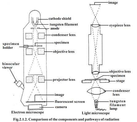
This microscope uses visible light for illuminating the object. It contains glass lenses that magnify the image of the object and focus the light on the retina of the observer’s eye. It has two lenses one at each end of a hollow tube. The lens closer to the object being viewed is called objective lens. The lens closer to the eye is called ocular lens or eyepiece. The object is illuminated by light beneath it. A third lens called condenser lens is located between the object and the light source and it serve to focus the light on the object.
Dark field microscope
This type of microscope is useful for viewing suspensions of bacteria. It has a special condenser that allows only rays of light scattered by structures within specimen. The result is an image that appears bright against a dark background, with a high degree of contrast. The process is similar to seeing dust particles floating in a sunbeam.
Phase contrast microscope
The phase contrast microscope has special fitments to the objective lens and sub stage condenser, the effect of which is to exaggerate the structural differences between the cell components. As a consequence, the structures within living, unstained cells become visible in high contrast and with good resolution. Phase contrast microscopy avoids the need to kill cells or to add dye to a specimen before it is observed microscopically.
Oil - immersion microscopy
In oil-immersion microscopy the light gathering properties of the objective lens are enhanced by placing oil in the space between the slide and objective lens. Normally the technique is used to view permanently mounted specimens.
The oil immersion lens gives higher magnification than the normal high- power objective lens.
Electron microscopy.
The electron microscopy uses the much shorter wavelengths of electrons to achieve resolutions as low as 3A°. Electromagnetic coils (i.e., magnetic lenses) are used to control and focus a beam of electrons accelerated from a heated metal wire by high voltages, in the range of 20,000 to 100,000 volts. The electrons are made to pass through the specimen. Electrons that do passes out of the object are focused by an objective coil (‘lens’). Finally a magnified image is produced by a projector coil or ‘lens’. The final image is viewed on a screen or is recorded on photographic film to produce electron micrograph. This type of electron microscope is called transmission electron microscope (TEM)
In a compound light microscope, the image is formed due to differences in light absorption. The electron microscope forms images as a result of differences in the way electrons are scattered by various regions of the object.

The degree to which electrons are scattered is determined by the thickness and atomic density of the object. Hence the specimens used in electron microscopy must be extremely thin. Living cells which are wet cannot be viewed in electron microscope.
Scanning electron microscopy (SEM)
This microscope has less resolution power than the TEM (i.e., about 200A°). However it is a very effective tool to study the surface topography of a specimen. The whole specimen is scanned by a beam of electrons. An image is created by the electrons reflected from the surface of the specimen. Scanning electron micrographs show depth of focus and a three dimensional image of the object.
References
T. Sargunam Stephen, Biology (Zoology). First Edition – 2005, Government of Tamilnadu.


|
|
|
|
دراسة يابانية لتقليل مخاطر أمراض المواليد منخفضي الوزن
|
|
|
|
|
|
|
اكتشاف أكبر مرجان في العالم قبالة سواحل جزر سليمان
|
|
|
|
|
|
|
اتحاد كليات الطب الملكية البريطانية يشيد بالمستوى العلمي لطلبة جامعة العميد وبيئتها التعليمية
|
|
|