

 النبات
 الحيوان
 الأحياء المجهرية
 علم الأمراض
 التقانة الإحيائية
 التقنية الحيوية المكروبية
 التقنية الحياتية النانوية
 علم الأجنة
 الأحياء الجزيئي
 علم وظائف الأعضاء
 الغدد
الغدد
 المضادات الحيوية
|
Read More
Date: 4-8-2016
Date: 2-8-2016
Date: 1-8-2016
|
Lacrimal Gland
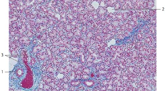
The lacrimal gland has approximately the shape of an almond. It consists of lobes that are intersected by connective tissue septae. The lacrimal gland is a tubular, purely serous gland without intercalate d ducts. The gland tubules end in intralobular ducts 1 , which merge to larger ducts. About 8–12 ducts channel the tears to the fornix conjunctivae. In cross-sections, the acini of the lacrimal gland resemble those of the parotid glands. They show all the morphological criteria of serous glands. The secretory cells of ten show a fine cytoplasmic granulation. The interstitial connective tissue is sometimes rich in lymphocytes and plasma cells. With increasing age, there are often also adipocytes 2 .
1 Intralobular ducts
2 Adipocytes
3 Artery
Stain: azan; magnification: × 80

Lacrimal Gland
The tubules of the lacrimal gland of ten have wide lumina. They are therefore often referred to as tubuloalveolar glands .The irregularly shaped tubules can be clearly assessed at higher magnification. Note the shape of the gland cells (secretor y cells). The round nuclei are in basal position. The cytoplasm appears light, and cell borders can be clearly recognized in some places. Myoepithelial cells are found between the gland epithelium and the basal membrane. Note the delicate connective tissue between the tubules (here stained blue). The secretory product of the lacrimal glands (tears) moisturizes the cornea and the conjunctiva of the eyeball as well as the eyelids.
Stain: azan; magnification: × 200

Lacrimal Gland
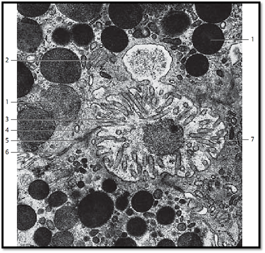
The varieties of secretory products of the exocrine and endocrine gland cells are store d in the cells as secretory granules or secretor y droplets. Intracellularly stored secretory granules may display a variety of appearances. The figure shows three acinar cells of the lacrimal gland. The secretory granules appear either homogeneous 1 or show low electron microscopic densities. They contain finely dispersed secretory particles. As shown in the figure, the secretory granules are released individually in response to stimuli. Microvilli 3 protrude into the acinar lumen. Note the granule in the lumen 7 . It has al-ready lost part of its membrane. The lateral surfaces of the secretory cells show apical junctional complexes (terminal bars) 456 .
1 Secretor y granules
2 Mitochondrion
3 Microvilli
4 Zonula occludens
5 Zonula adherens
6 Desmosome
7 Secretory granule in the acinar lumen
Electron microscopy; magnification: × 25 000

References
Kuehnel, W.(2003). Color Atlas of Cytology, Histology, and Microscopic Anatomy. 4th edition . Institute of Anatomy Universitätzu Luebeck Luebeck, Germany . Thieme Stuttgart · New York .


|
|
|
|
تفوقت في الاختبار على الجميع.. فاكهة "خارقة" في عالم التغذية
|
|
|
|
|
|
|
أمين عام أوبك: النفط الخام والغاز الطبيعي "هبة من الله"
|
|
|
|
|
|
|
قسم شؤون المعارف ينظم دورة عن آليات عمل الفهارس الفنية للموسوعات والكتب لملاكاته
|
|
|