

 النبات
 الحيوان
 الأحياء المجهرية
 علم الأمراض
 التقانة الإحيائية
 التقنية الحيوية المكروبية
 التقنية الحياتية النانوية
 علم الأجنة
 الأحياء الجزيئي
 علم وظائف الأعضاء
 الغدد
الغدد
 المضادات الحيوية
|
Read More
Date: 28-12-2020
Date: 1-1-2021
Date: 2-8-2016
|
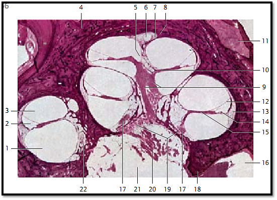
Inner Ear-Cochlea
Longitudinal section of a human cochlea.
1 Scala tympani (contains perilymph)
2 Cochlear duct (contains endolymph), triangular in cross-sections
3 Scala vestibuli (contains perilymph)
4 Osseous spiral lamina
5 Helicotrema (scala vestibuli and scala tympani join)
6 Cecum cupulare (copular blind sac, caecum cupulare)
7 Modiolus (end of the osseous spiral lamina)
8 Cochlear cupola (azimuth of the apical cochlear turn)
9 Longitudinal canals of the modioli (canales longitudinales modioli), central inner canals of the cochlea
10 Spiral canal of the modioli (canalis spiralis modioli), canal in the outer wall of the osseous spiral lamina
11 Facial nerve in the bony facial canal
12 Vestibular membrane (Reissner’s membrane, membrana vestibularis, upper wall of the cochlea)
13 Spiral crest, spiral ligament (ligamentum spirale cochlea)
14 Basilar membrane (connective tissue between cochlear duct and scala tympani)
15 Osseous spiral lamina
16 Area of the facial nerve of the internal acoustic meatus (fundus)
17 Cochlear ganglion (cochlear nerve)
18 Transverse crest
19 Base of the modiolus (base of the cochlea)
20 Cochlear ner ve
21 Internal acoustic meatus (fundus)
22 Spiral canal of the modioli Stain: hematoxylin-eosin; magnification: × 10

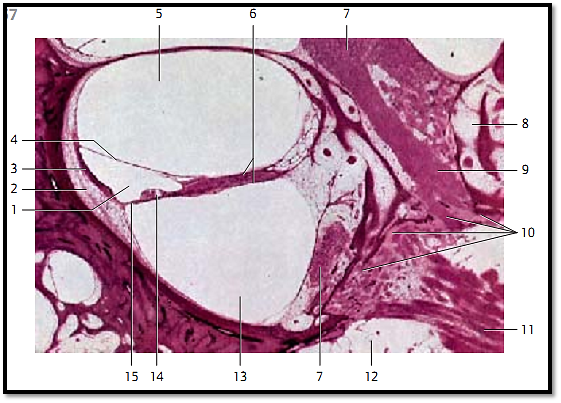
Cochlea—Inner Ear
Middle b end of the cochlea.
1 Cochlear duct (contains endolymph)
2 Spiral ligament, spiral crest
3 Stria vascularis, vascularized tissue over the spiral prominence
4 Reissner’s membrane, vestibular membrane
5 Scala vestibuli
6 Osseous spiral lamina
7 Cochlear ganglion
8 Spiral canal of the modioli (bony canal in the outer wall of the osseous spiral lamina)
9 Longitudinal canal of the modioli (central inner canal)
10 Tractus spiralis foraminosus, a few spirally arranged openings around the inner cochlear
canal, which permit access of cochlear nerve ganglia to the cochlea
11 Cochlear nerve (branch of the vestibulocochlear nerve for the cochlear audiosensory organ)
12 Internal acoustic meatus (fundus)
13 Scala tympani (contains perilymph)
14 Organ of Corti
15 Basilar membrane (connective tissue layer between cochlear duct and scala tympani)
Stain: hematoxylin-eosin; magnification: × 25

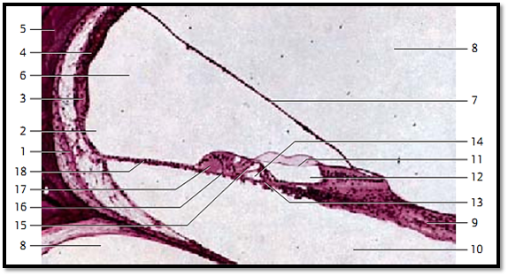
Cochlea—Inner Ear
Section of the cochlear duct from the apical turn of the cochlea with theorgan of Corti ( organum spirale).
1 Spiral ligament, spiral crest
2 External spiral groove of the cochlear duct
3 Spiral prominence (border of the external spiral groove)
4 Stria vascularis
5 Bony wall of the cochlea
6 Cochlear duct
7 Vestibular membrane, Reissner’s membrane
8 Scala vestibuli
9 Cochlear nerve
10 Scala tympani
11 Tectorial membrane (located over the organ of Corti and the inner cochlear canal)
12 Inner cochlear canal (canalis spiralis cochleae)
13 Inner hair cells
14 Inner tunnel, contains endolymph
15 Outer hair cells
16 Cells of Henson
17 Cells of Claudius
18 Basilar membrane
Stain: hematoxylin-eosin; magnification: × 80

References
Kuehnel, W.(2003). Color Atlas of Cytology, Histology, and Microscopic Anatomy. 4th edition . Institute of Anatomy Universitätzu Luebeck Luebeck, Germany . Thieme Stuttgart · New York .


|
|
|
|
دخلت غرفة فنسيت ماذا تريد من داخلها.. خبير يفسر الحالة
|
|
|
|
|
|
|
ثورة طبية.. ابتكار أصغر جهاز لتنظيم ضربات القلب في العالم
|
|
|
|
|
|
|
العتبة العباسية المقدسة تقدم دعوة إلى كلية مزايا الجامعة للمشاركة في حفل التخرج المركزي الخامس
|
|
|