

 النبات
 الحيوان
 الأحياء المجهرية
 علم الأمراض
 التقانة الإحيائية
 التقنية الحيوية المكروبية
 التقنية الحياتية النانوية
 علم الأجنة
 الأحياء الجزيئي
 علم وظائف الأعضاء
 الغدد
الغدد
 المضادات الحيوية
|
Read More
Date: 11-1-2017
Date: 31-7-2016
Date: 4-8-2016
|
Peroxisomes
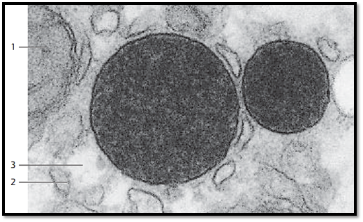
In 1965, C. de Duve discovered peroxisomes in liver cells. J. A . Rhodin had previously described them as “microbodies.” They are small, spherical organelles with a diameter of about 0.2–1.5 μmm, which are ubiquitous in cells. Peroxisomes have a single membrane as their outer border. Their finely granular or homogeneous content is electron-dense. Occasionally, peroxisomes enclose paracrystalline materials. Peroxisomes are respiratory organelles. They contain various oxidases, catalase and the enzymes for the β-oxidation of fatty acids. Peroxisomes owe their name to their peroxidative enzyme content. They play an important role in the detoxification of cells. Example: the enzyme catalase will split hydrogen peroxide, a lethal cell poison. Genetic diseases that are based on peroxisomal defects include Zellweger syndrome , Ref sum syndrome and adrenoleukodystrophy . Section from an epithelial cell (human liver) with two peroxisomes of different sizes.
1 Section through a mitochondrion
2 Smooth endoplasmic reticulum (sER) membranes
3 Areas with cellular glycogen (glycogen removed)

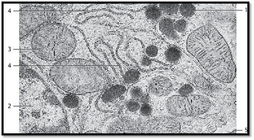
Section through a mouse epithelial cell from a proximal kidney tubule with several peroxisomes, which are rich in catalase 1 .
1 Peroxisomes
2 Nucleus (partial section)
3 Mitochondria (crista-type)
4 Granular ER (rER) membranes
5 Peritubular connective tissue space

References
Kuehnel, W.(2003). Color Atlas of Cytology, Histology, and Microscopic Anatomy. 4th edition . Institute of Anatomy Universitätzu Luebeck Luebeck, Germany . Thieme Stuttgart · New York .


|
|
|
|
دخلت غرفة فنسيت ماذا تريد من داخلها.. خبير يفسر الحالة
|
|
|
|
|
|
|
ثورة طبية.. ابتكار أصغر جهاز لتنظيم ضربات القلب في العالم
|
|
|
|
|
|
|
سماحة السيد الصافي يؤكد ضرورة تعريف المجتمعات بأهمية مبادئ أهل البيت (عليهم السلام) في إيجاد حلول للمشاكل الاجتماعية
|
|
|