آخر المواضيع المضافة

 النبات
الحيوان
الأحياء المجهرية
علم الأمراض
التقانة الإحيائية
التقنية الحيوية المكروبية
التقنية الحياتية النانوية
علم الأجنة
الأحياء الجزيئي
علم وظائف الأعضاء
الغدد
المضادات الحيوية
النبات
الحيوان
الأحياء المجهرية
علم الأمراض
التقانة الإحيائية
التقنية الحيوية المكروبية
التقنية الحياتية النانوية
علم الأجنة
الأحياء الجزيئي
علم وظائف الأعضاء
الغدد
المضادات الحيوية| Smooth Muscle-Artery |


|
|
Read More
Date: 26-7-2016
Date: 18-1-2017
Date: 3-1-2017
|
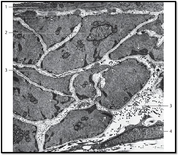
Smooth Muscle-Artery
This figure shows a cross-section through the smooth muscle cells of the tunica me dia (vascular coat) of the parotid gland. The cell at the top center underneath the endothelium 1 contains a nucleus 2 . In addition, there are small groups of ribosomes, a few crista-type mitochondria and groups or rows of vesicles, called caveolae , and pre dominantly located along the sarcolemma, small dense areas (dense spots ) , which serve as focal adhesion points for contractile fibers. The myofilaments in this figure are cut across their long axis, and therefore appear as small spots in the cytoplasm. Nexuses (contacts) are present in a few places between neighboring muscle cells . They are loci with low electrical resistance and serve as excitation conduits. Note that a basal membrane envelops the muscle cells. The extracellular collagen fibrils have been pre dominantly cut across their axis .
1 Endothelium
2 Nucleus
3 Extracellular connective tissue space with collagen fibrils
4 Fibrocyte
Electron microscopy; magnification: × 9500
References
Kuehnel, W.(2003). Color Atlas of Cytology, Histology, and Microscopic Anatomy. 4th edition . Institute of Anatomy Universitätzu Luebeck Luebeck, Germany . Thieme Stuttgart · New York .

|
|
|
|
دخلت غرفة فنسيت ماذا تريد من داخلها.. خبير يفسر الحالة
|
|
|
|
|
|
|
ثورة طبية.. ابتكار أصغر جهاز لتنظيم ضربات القلب في العالم
|
|
|
|
|
|
|
العتبة العباسية المقدسة تدعو جامعة ميسان للمشاركة في الحفل المركزي لتخرج طلبة الجامعات
|
|
|