

 النبات
 الحيوان
 الأحياء المجهرية
 علم الأمراض
 التقانة الإحيائية
 التقنية الحيوية المكروبية
 التقنية الحياتية النانوية
 علم الأجنة
 الأحياء الجزيئي
 علم وظائف الأعضاء
 الغدد
الغدد
 المضادات الحيوية
|
Read More
Date: 14-8-2016
Date: 27-7-2016
Date: 25-7-2016
|
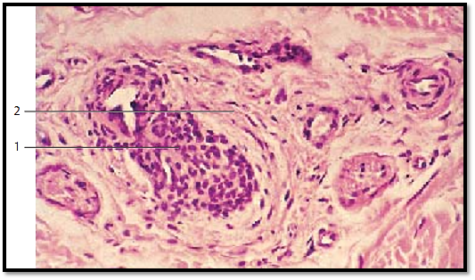
Arteriovenous Anastomoses
The connection between the very small arteries and venules is not always made via the capillary network but in many cases, via immediate contractile shunts. These anastomosis vessels reroute the bloodstream directly from the arterial high-pressure circulation to the venous low-pressure system, thus circumventing the capillary network . In humans, arteriovenous anastomosis vessels occur in large numbers in the extremities. They are known as Masson’s organs or Hoyer - Grosser organs (glomus neuromyoarteriale, anastomosis arteriovenosa glomiformis). These organs show specific forms of epithelial myocytes. Longitudinal muscle bundles already exist on the arterioles where the shunts originate. The connection between artery and vein consists of winding tubules 1 with thick walls (about 40–60 μm) and narrow lumina (10–30 μm). Unmyelinated nerve cells of ten weave around these looping coils of vessels. A layer of connective tissue separates the shunt vessels and nerve cells from the surrounding tissue. This figure shows such a shunt structure. The presence of a strong layer of epithelioid muscle cells under the epithelial layer emphasizes the shunts. Epithelioid muscle cells are fibril-free. Their cytoplasm is only slightly stained with the procedure used here. The by-pass canals (shunts) do not have an internal elastic membrane (tunica elastica interna). Human fingertip.
Stain: hematoxylin-eosin; magnification: × 200

References
Kuehnel, W.(2003). Color Atlas of Cytology, Histology, and Microscopic Anatomy. 4th edition . Institute of Anatomy Universitätzu Luebeck Luebeck, Germany . Thieme Stuttgart · New York .


|
|
|
|
علامات بسيطة في جسدك قد تنذر بمرض "قاتل"
|
|
|
|
|
|
|
أول صور ثلاثية الأبعاد للغدة الزعترية البشرية
|
|
|
|
|
|
|
مكتبة أمّ البنين النسويّة تصدر العدد 212 من مجلّة رياض الزهراء (عليها السلام)
|
|
|