

 النبات
 الحيوان
 الأحياء المجهرية
 علم الأمراض
 التقانة الإحيائية
 التقنية الحيوية المكروبية
 التقنية الحياتية النانوية
 علم الأجنة
 الأحياء الجزيئي
 علم وظائف الأعضاء
 الغدد
الغدد
 المضادات الحيوية
|
Read More
Date: 23-1-2017
Date: 4-1-2017
Date: 15-1-2017
|
Extraepithelial Glands—Serous Glands
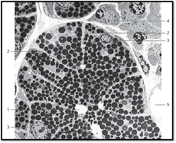
The secretory cells of active glands accumulate visible supplies of secretory products. This leads to enrichment in secretory materials and their precursors. In the process, the numb er of conspicuous secretor y granules or secretory droplets increases. The acinar cells of the parotid gland show the typical attributes of serous glands. Their many secretor y granules are distribute d over the entire cell. In electron microscopy, they appear as osmiophilic spheres. The electron densities of granular membrane and contents are the same. The granular membrane is therefore hardly visible, even at high magnification. The secretory granules are generally released into the lumen 1 as single granules (exocytosis). However, occasionally the secretory granules in serous gland cells may fuse before they are extruded. This figure shows an acinus of the parotid gland with osmiophilic secretory granules. On average, the gland cells are cone-shaped. There are secretor y canaliculi 2 between gland cells. The nuclei 3 are locate d in the basal space.
1 Acinar lumen
2 Intercellular secretor y canaliculi
3 Cell nuclei
4 Connective tissue cells
5 Capillary
Electron microscopy; magnification: × 1800

References
Kuehnel, W.(2003). Color Atlas of Cytology, Histology, and Microscopic Anatomy. 4th edition . Institute of Anatomy Universitätzu Luebeck Luebeck, Germany . Thieme Stuttgar t · New York .


|
|
|
|
"إنقاص الوزن".. مشروب تقليدي قد يتفوق على حقن "أوزيمبيك"
|
|
|
|
|
|
|
الصين تحقق اختراقا بطائرة مسيرة مزودة بالذكاء الاصطناعي
|
|
|
|
|
|
|
قسم شؤون المعارف ووفد من جامعة البصرة يبحثان سبل تعزيز التعاون المشترك
|
|
|