

 النبات
 الحيوان
 الأحياء المجهرية
 علم الأمراض
 التقانة الإحيائية
 التقنية الحيوية المكروبية
 التقنية الحياتية النانوية
 علم الأجنة
 الأحياء الجزيئي
 علم وظائف الأعضاء
 الغدد
الغدد
 المضادات الحيوية
|
Read More
Date: 9-6-2021
Date: 31-3-2021
Date: 14-11-2020
|
Avidin-Biotin System
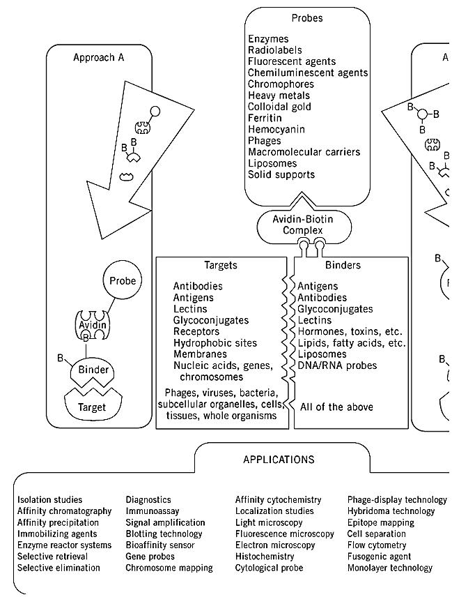
The avidin-biotin system has become one of the major mainstays in biochemical analysis and has far-reaching application in biotechnology, industry, and clinical medicine (1). The general idea of the avidin-biotin system is that biotin, a low molecular weight vitamin, can be chemically coupled to other low or high molecular weight molecules (e.g., proteins, hormones, DNA molecules, etc.). The biotin moiety is still recognized by avidin or streptavidin, either as the native protein or in derivatized form containing any one of a number of reporter groups or probes. The principle of this system is illustrated in Fig. 1.

Figure 1. Overview of the avidin-biotin system and the two major strategies for the various applications. In both approach desired experimental system is combined with a biotinylated binder molecule. Approach A involves direct interaction with probe. In approach B, avidin is a sandwich between the biotinylated binder and the biotinylated probe. Various targets, b the applications are listed.
The avidin-biotin system has become a “universal” tool in most of the fields of the biological sciences, because of studies that commenced in the mid 1970s and the constant development that has continued until today (2-6). The system has been applied for a wide variety of purposes (Fig. 1) and has recently been adapted for clinical use for the localization, imaging, and therapy of cancer (7). The binder and the target can be any of the components listed in Figure 1. What is required for this system is the capacity to biotinylate a binding entity so that the specificity and activity of the binding function is retained. For different approaches to biotinylation and the reagents used for binding to different functional groups. As can be seen, most of the functional groups on biological molecules can be modified with biotin. Because of its charge neutrality and lack of glycosylation, streptavidin is generally preferred over egg-white avidin in many applications, although new derivatives of avidin (e.g., Neutravidin) may prove advantageous and less expensive. The final component added to the system is the probe. The various probes and their potential uses are shown in Fig. 1. The probes are prepared in two ways. They are chemically conjugated directly to avidin or streptavidin (Fig. 1, Approach A) and are fluorescent, radioactive, or other types of macromolecules (proteins, polysaccharides, etc.). A second approach is to biotinylate the probes and to interact them with streptavidin under subsaturating ratios, thus leaving extra binding sites vacant (Fig. 1, Approach B). More recently, fusion proteins have been prepared of streptavidin with different enzymes and native fluorescent proteins.
In many cases, such as affinity chromatographic applications, more reversible binding of biotin to avidin would be a distinct advantage. In this context, streptavidin has a critical disadvantage in that the interaction between its subunits is very strong, and it cannot be used when reversibility of the interaction is desired. This is in contrast to avidin, from which an immobilized monovalent form can be produced (8), and the immobilized avidin monomer binds biotinylated compounds reversibly (9).
Site-directed mutagenesis and chemical modification studies are currently being performed on both avidin and streptavidin to understand better their interaction with biotin, the interaction between their subunits, and, perhaps eventually, for better application in the previously-mentioned systems (10 ,11)In this regard, the single, critical tyrosine residue of the binding sites of avidin and streptavidin was nitrated, and the tetrameric structures of the resultant “nitro avidin” and “nitro streptavidin” were retained (12). The biotin-binding property also had sufficiently high affinity for a variety of applications, including affinity chromatography, enzyme immobilization, and phage-display technology (13, 14). The major difference between the nitro avidins and the native molecules is that biotinylated compounds are released by competition with free biotin, and then the latter is liberated by treating the column with basic solutions (pH 10), thereby regenerating the original biotin-binding capacity of the nitro avidin affinity column. Reduced or altered binding characteristics are also conferred on the binding sites of avidin or streptavidin by site-directed mutagenesis of selected binding site residues, such as tryptophans.
References
1. M. Wilchek and E. A. Bayer, eds. (1990) Avidin-Biotin Technology Methods in Enzymology, Vol. 184, Academic Press, San Diego.
2. E. A. Bayer and M. Wilchek (1978) Trends Biochem. Sci. 3, N237–N239.
3. E. A. Bayer and M. Wilchek (1980) Methods Biochem. Anal. 26, 1–45.
4. M. Wilchek and E. A. Bayer (1984) Immunol. Today 5, 39–43.
5. M. Wilchek and E. A. Bayer (1989) In Protein Recognition of Immobilized Ligands (T. W. Hutchens, ed.), Alan R. Liss, New York, pp. 83–90.
6. E. A. Bayer and M. Wilchek (1996) In Immunoassay (E. P. Diamandis and T. K. Christopoulos, eds.), Academic Press, San Diego, pp. 237–267. 7. G. Paganelli, P. Magnani, A. G. Siccardi, and F. Fazio (1995) In Cancer Therapy with Radiolabeled Antibodies (D. M. Goldenberg, ed.), CRC Press, Boca Raton, FL, pp. 239–254.
8. N. M. Green and E. J. Toms (1973) Biochem. J. 133, 687–700.
9. K. P. Henrikson, S. H. G. Allen, and W. L. Maloy (1979) Anal. Biochem. 94, 366–370.
10. A. Chilkoti, P. H. Tan, and P. S. Stayton (1995) Proc. Natl. Acad. Sci. USA 92, 1754–1758.
11. A. Chilkoti, B. L. Schwartz, R. D. Smith, C. J. Long, and P. S. Stayton (1995) Bio/Technology 13, 1198-1204 .
12. E. Morag, E. A. Bayer, and M. Wilchek (1996) Biochem. J. 316, 193–199.
13. E. Morag, E. A. Bayer, and M. Wilchek (1996) Anal. Biochem. 243, 257–263.
14. M. E. M. Balass, E. A. Bayer, S. Fuchs, M. Wilchek, and E. Katchalski-Katzir (1996) Anal. Biochem. 243, 264–269.


|
|
|
|
علامات بسيطة في جسدك قد تنذر بمرض "قاتل"
|
|
|
|
|
|
|
أول صور ثلاثية الأبعاد للغدة الزعترية البشرية
|
|
|
|
|
|
|
مكتبة أمّ البنين النسويّة تصدر العدد 212 من مجلّة رياض الزهراء (عليها السلام)
|
|
|