

 النبات
 الحيوان
 الأحياء المجهرية
 علم الأمراض
 التقانة الإحيائية
 التقنية الحيوية المكروبية
 التقنية الحياتية النانوية
 علم الأجنة
 الأحياء الجزيئي
 علم وظائف الأعضاء
 الغدد
الغدد
 المضادات الحيوية
|
Read More
Date: 6-12-2015
Date: 31-5-2021
Date: 16-12-2015
|
Autoradiography
Autoradiography is the technique of recording an image of a preparation containing beta-particle emitting radioactivity, using photographic film, X-ray-sensitive film, an emulsion, or other radiation-sensitive medium. The samples are placed directly against the film for a period of time to allow radioactive emissions from the sample to interact with the film emulsion and create an image. A photographic emulsion is a suspension of crystals of silver bromide embedded in gelatin. When crystals of silver bromide are struck by charged-particle or photon radiation, the silver atoms are ionized and form an invisible latent image. After exposure to the sample, the photographic grains in the emulsion are fixed using standard photographic developing, which removes silver bromide that has not been ionized. After the emulsion is developed, each small aggregate of reduced silver atoms becomes a visible dark spot on the emulsion; collectively, they make up the photographic image.
Production of a visible silver grain requires a number of ionization events, so the photographic response is not exactly linear to the amount of radiation present. Preflashing the film with a uniform low intensity of light “primes” each grain of silver to become reduced and visible after absorbing just one or a very few additional beta particles from the sample. This increases substantially the sensitivity of the film and also makes the photographic response more directly proportional to the amount of radiation in the sample. The signal-to-noise ratio is often increased by exposing the film to low temperatures. The sensitivity can also be enhanced by using scintillation screens that emit visible light on encountering a beta particle; the light is recorded by the film.
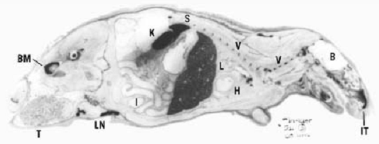
Autoradiography has a large number of practical applications in the biological, chemical, and physical sciences, because it provides both qualitative and quantitative information (eg., images and amounts present). It may be used to image large, small, and microscopic specimens, including sectioned whole organisms, organs, tissues, cellular structures, and nucleic acids that contain some radiolabeled compound. An example of a whole-animal autoradiograph is shown in Figure 1. Cells
may be autoradiographed either in culture as a monolayer, on a glass slide, or on thinly sectioned living tissues from an animal organ or tumor. Microautoradioagraphy involves coating the sample directly with a radiation-sensitive emulsion; cellular constituents that have incorporated the radiolabel can be clearly identified. Autoradiography is used with electrophoresis or chromatography to image radiolabeled macromolecules and other separated chemicals for quantitative analysis. For example, autoradiography is useful for indicating the position of hybridized nucleic acids on Southern blots and Northern blots, and of proteins on Western blots.

Figure 1. Whole-body autoradiograph of a rat that had been injected with indium-111 chloride. Courtesy of B. Anders Jönsson, Lund University, Sweden.
1.Historical Development
The first autoradiograph, or autoradiogram, was made in 1859 by Crookes, who placed uranium rocks on photographic plates (1). Crookes would not understand the process by which the images were created until 37 years later when he visited the laboratory of Henri Becquerel. Bequerel discovered gamma rays from naturally radioactive uranium salts in 1896 when he placed uranium together with photographic plates that had been placed inside black paper to protect them from sunlight (1). This discovery closely followed the discovery of X rays by Wilhelm Roentgen in 1895 using a Crookes electron tube.
The French photographer Nicéphore Niepce had observed the darkening of silver grains on photographic plates by uranium in 1867, but he did not recognize any practical applications of this phenomenon (2). Although George de Hevesy, the pioneer of radioactive tracers, had used bismuth isotopes in animals in 1923, the first autoradiographs of radioactively labeled animal tissues were made in 1924 by Lacassagne in France (1-3). Lacassagne fed polonium-210 to rabbits and later placed thin sections of rabbit kidney tissues in paraffin blocks against photographic plates. Gettler made the first autoradiographs of human tissues from deceased persons who had ingested radium chloride in 1933 (1).
The earliest images were of poor quality. Autoradiographic techniques were improved by Leblond, who, in 1943, showed the microdistribution of iodine-131 in cells (1, 2). In 1946, Bélanger and Leblond (4) developed a method for locating radioactive elements in tissues by covering histological sections with a photographic emulsion. In 1954, Gabriel introduced techniques for identifying radioiodine using human serum albumin zone paper electrophoresis (1). In 1953, techniques were developed for labeling nucleic acids and studying kinetics of cell mitosis and division (5). High resolution techniques have more recently been employed for study of other metabolic and pharmacokinetic processes in molecular biology.
2. Film-Less Autoradiography
Modern trends in autoradiography involve replacing high speed X-ray film with radiation detector systems, laser scanners, and computer-based imaging systems. A variety of radiation-detecting crystals and phosphors have been developed for this purpose. Storage phosphor screens are more sensitive, by a factor of about 20–100 for beta-emitting radionuclides (2), and they are reusable. The exposure time is also much less, by a factor of about 10, over conventional X-ray film, and samples may be processed at room temperature and without a darkroom or chemicals for film developing.
Applications of filmless autoradiography include two-dimensional gels, Southern blots, Northern blots, immunoblots, and quantitative polymerase chain reaction (PCR) (2).
Microchannel array detectors have been introduced to replace both X-ray films and phosphor screens (8) .The new instruments are faster (by a factor of ~10) and have greater image resolution than do phosphor screens for detecting latent images from hybridization studies using macromolecules labeled with carbon-14, sulfur-35, phosphorus-32, and iodine-125 from flat gels, blots, membranes, tissue slices, and other flat specimens.
References
.1M. Brucer (1990) A Chronology of Nuclear Medicine, Heritage Publications Inc., St. Louis, MO.
2. R. Wegmann, N. Balmain, S. Ricard-Blum, and S. Guha (1995) Cell. Mol. Biol. 41, 1–20.
3. A. Lacassagne, J. Lattes, and J. Lavedan (1925) J. Radiol. Electr. 9, 1–14.
4. L.-F. Bélanger and C. P. Leblond (1946) Endocrinology 39, 386–400.
5. A. Howard and S. R. Pelc (1970) Heredity 6 (suppl.), 261–273.
6. J. G. Gall and M. Pardue (1969) Proc. Natl. Acad. Sci. USA 63, 378–383.
7. E. M. Southern (1975) J. Mol. Biol. 98, 503–517.
8. Packard Instrument Company (1993) Enter the New Era of Instant Autoradiography: The InstantImager™, promotional literature from Packard Instrument Company, Meriden, Conn.


|
|
|
|
علامات بسيطة في جسدك قد تنذر بمرض "قاتل"
|
|
|
|
|
|
|
أول صور ثلاثية الأبعاد للغدة الزعترية البشرية
|
|
|
|
|
|
|
مكتبة أمّ البنين النسويّة تصدر العدد 212 من مجلّة رياض الزهراء (عليها السلام)
|
|
|