


 النبات
 الحيوان
 الأحياء المجهرية
 علم الأمراض
 التقانة الإحيائية
 التقنية الحيوية المكروبية
 التقنية الحياتية النانوية
 علم الأجنة
 الأحياء الجزيئي
 علم وظائف الأعضاء
 الغدد
الغدد
 المضادات الحيوية
|
Read More
Date: 2-11-2015
Date: 5-11-2015
Date: 30-10-2015
|
Developmental Biology
The process of sexual reproduction ensures the formation of a diploid zygote which could constitute the next generation. A zygote is a single celled structure. By an ontogenetic process the zygote undergoes various developmental phases resulting in multicellular embryonic organization. These phases include cleavage, gastrulation, neurulation, organogenesis and the period of growth and histological differentiation. Inspite of the fact that organisms vary in their structure, form and mode of life, the processes of embryogenesis, development and differention are remarkably similar in all metazoans. Till later stages of development a fundamental uniform pattern in development can be observed. The ontogenetic stages also reflect the historical development of species or phylogenetic development.
Realising the mode of formation of a young individual of the next generation has always interested human mind. There is a recorded history of human natural curiosity in sexual reproduction from very early period. The ‘Susruta samhita’, a monumental Indian medical book, written during second or third century A.D., describes the development of a human child in the mother’s womb.
The earliest recorded work had been done by Aristotle (384-322 BC). His classical work De Generatione Animalium is concerned with the generation of animals. It describes the reproduction and development of many kinds of animals. In his another work “De Historia Animalaium, Aristotle provides an account of the development of the hen’s egg. He compared reproductive methods of different animals and provided a classification based on that. By observing the development of hen’s egg he concluded that the development always proceeds from simple formless beginning to the complex organization of the adult. For this speculative idea he provided the name epigenesis. Through his remarkable observation and speculations Aritotle established ‘embryology’ as an independent field in science. Thus to-day he is regarded as the founder of the science of embryology.
After the period of early Greek thinkers this discipline once again got the attention of the scientists from the beginning of the 17th century. Through the contributions made by various workers like Von Baer, E. Haeckel, O. Hertwig, E.B Wilson, Spemann, C.M Child, Maclean and others rapid advancements were being made in the understanding of developmental processes in animals. Modem embryology has utilized all tools made available from other branches of science and diversified into branches such as ‘Experimental embryology’, ‘Chemical embryology’, ‘Comparative embryology’ and Descriptive embryology. Such studies have paved the way for meeting the challenges of to-day’s world through works on cloning techniques, tissue culture, stem cell researches, ‘in vitro’ fertilization, organ transplantations, regeneration, tissue grafting and other medical and non-medical fields.
Gametogenesis:
The process of embryonic development in sexually reproducing multicellular organisms is made possible through processes of gametogenesis and fertilization. Gametogenesis is the formation of sex cells or reproductive cells or gametes. It happens in primary sex organs called gonads. The male and female gonads, namely the testis and ovary contain primordial germ cells. These cells are responsible for the production of gametes.
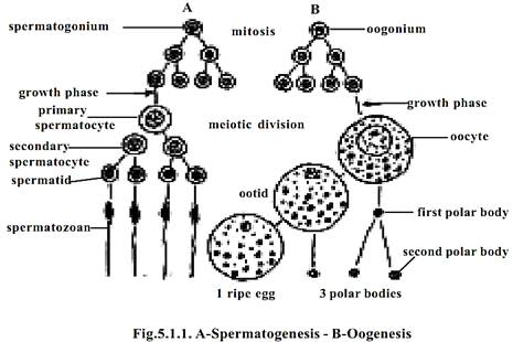
Spermatogenesis:
In the testis of vertebrates the specialized tissue for the process of spermatogenesis are located in the seminiferous tubules. The primordial germ cells of these tubules produce cells which ultimately become sperm mother cells or spermatogonia. Through a growth phase the speramtogonia get converted into primary spermatocytes. These are diploid cells. They undergo meiotic cell division. Initially the I Meiosis results in the formation of secondary spermatocytes. Through II Meiosis they form spermatids. The spermatids are haploid in nature. By a process of spermiogenesis or spermioteliosis they get differentiated into specialized cells called spermatozoa.
Oogenesis:
A similar process happens inside the female gonad, namely the ovary for the production of Ova. This process that happens in the primordial germ cell of the ovary passes through stages of primary oogonia, primary oocyte and secondary oocyte. These stages are conducted by meiotic cell divisions. Thus the final product, namely the ovum is a haploid female reproductive cell.
Fertilization:
Embryogenesis could occur only after the fertilization of the ovum or egg. Fertilization provides the diploid nature to the cell. Thus all the somatic cells of the embryo will remain diploid. Further, the process of fertilization triggers or initiates the initial stages of embryogenesis. During the process of fertilization the sperm and ovum of the same species approach and come in contact with each other. The entry of sperm initiates further changes in the egg. The haploid nuclei of the sperm and ovum fuse, resulting in the formation of a diploid zygote nucleus. This process of nuclear fusion is known as syngamy or amphimixis.
Types of eggs
For the embryo to develop inside a fertilized egg nutrition is needed. The amount of food needed varies for different organisms. It normally depends on the duration of development. Food is provided in the form of yolk. It may be ‘fatty yolk’ or protein yolk’. It is provided by the ovary during differentiation of the egg. Due to accumulation of yolk a maturing egg rapidly increases in size. In amphibian eggs yolk occurs in the form of large granules, called yolk platelets. Chemically, the yolk platelets contain two main proteinaceous substances namely phosvitin and lipovitellin.
The amount of yolk is an important determining factor for further patterns in embryological stages. The amount of yolk influences cleavage and gastrulation methods.
The eggs can be classified based on amount and distribution of yolk.
Amount of yolk and egg types:
In certain animals the developmental stages are not very elaborate. The final ‘young one’ born may be very simple in structure and organization. Such conditions remain in animals like Hydra, Sea urchin, Amphioxus and Placental mammals. In the eggs of such organisms due to brevity of the growth period the amount of yolk is much reduced. Such eggs are said to be Microlecithal or oligolecithal.
In certain other animals the eggs need to release young ones in a more self-supportive condition. Hence for such eggs the amount of yolk is considerable in quantity. Such eggs with moderate amount of yolk are called mesolecithal eggs. Such eggs are produced by annelid worms, molluscs and amphibians.
In some animals the growth and differentiation of the embryo is much more elaborate. The growth period is sufficiently long. Hence for supporting the embryo in development the eggs contain large quantity of yolk. Such eggs are termed as Megalecithal or Macrolecithal eggs. The eggs of reptiles and birds are considered as macrolecithal. Further these eggs are covered by a calcareous shell. It is a protective structure for laying the eggs on lands. Such eggs are called cleidoic eggs or land eggs
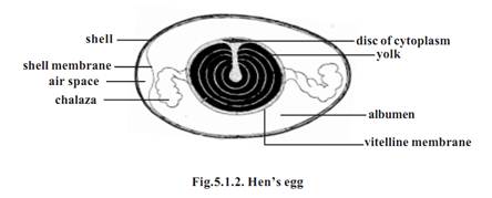
Distribution of yolk.
The pattern of cleavage and the consequent gastrulation processes are affected by the distribution of yolk within the egg. According to the pattern of dispersal of yolk the following egg types had been identified.
Homolecithal or isolecithal eggs
1-Eggs of this type have the yolk disbursed in the entire cytoplasm. The distribution is somewhat uniform in animal, vegetal poles and the equatorial

region. In such eggs the cleavage will be deeper and may bisect the eggs connecting the two poles. All microlecithal eggs have this nature.
2-Telolecithal eggs
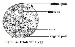
All eggs have polarity. In polarity, the eggs have an innate nature for to be differentiated into upper animal pole and lower vegetal pole. The polar nature is mainly due to the denser material in the cytoplasm, namely yolk. The yolk in the egg will normally get concentrated in the vegetal pole. The cytoplasm with the nucleus will occupy the upper animal pole. The extent of vegetal pole is determined by the amount of yolk. Thus the eggs having a polarized distribution of yolk in the cytoplasm are referred to as Telolecithal eggs. Mesolecithal and macrolecithal eggs remain as telolecithal eggs.
3-Centrolecithal eggs

An egg need not be spherical always. In invertebrate animals oval shaped eggs are seen. The pattern of cleavage and further gastrulation also deviate from that of the vertebrates. In insects the eggs are oval in shape and the yolk remains in the center of the egg. However, the eggs of echinoderms are similar to that of the vertebrates.
Cleavage and types - Frog’s egg
The process of cleavage remains one of the earliest mechanical activities in the conversion of a single celled egg into a multicellular embryo. It is initiated by the sperm during fertilization. However in parthenogenetic eggs cleavage can commence without the influence of fertilization.
The process of cleavage or cellulation happens through repeated mitotic divisions. These divisions result in cells called blastomeres. In later stages of development the blastomeres occupy different regions and differentiate into several types of body cells.
The first cleavage of frog’s egg was observed by Swammerdam in 1738. The entire process of cleavage in frog’s egg was studied by Prevost and Dumas in 1824. With the development of microscopes cleavages and further stages were observed in the eggs of sea urchin, star fishes, amphioxus and hen’s eggs.
From all these studies it has become clear that all divisions in cleavage are mitotic. The mitotic process is very rapid. In the eggs of sea urchin division of the blastomeres can be observed every 30 minutes. As the cleavage progresses the resultant daughter cells, namely the blastomeres get reduced in size. During cleavage there is no growth in the blastomeres. The total size and volume of the embryo remains the same. The cleavages result in a compact mass of blastomeres called morula. It gets transformed into blastula. While the wall of the blastula is called the blastoderm, the central cavity is called the blastocoel.
The planes of cleavage
An egg can be divided from different planes during cleavage. Depending on the position of the cleavage furrow the planes of cleavage are named.
1-Meridional cleavage: The plane of cleavage lies on the animal vegetal axis. It bisects both the poles of the egg. Thus the egg is divided into two equal halves
2-Vertical cleavage: The cleavage furrows may lie on either side of the meridional plane. The furrows pass from animal to vegital pole. The cleaved cells may be unequal in size
3-Equatorial cleavage: This cleavage plane bisects the egg at right angles to the main axis. It lies on the equatorial plane. It divides the egg into two halves
4-Latitudinal cleavage: It is similar to the equatorial plane, but it lies on either side of the equator. It is also called as transverse or horizontal cleavage
Influence of yolk on cleavage
Yolk is needed for embryonic development. However the fertilized egg has to undergo all stages of development and result in a suitable ‘young form’ initiating next generation. Somehow with all the influences of yolk the developmental procedures are so adapted and modified that a well formed embryo will result. The initial influence of yolk is felt during the process of cleavage.
The amount of the yolk and its distribution affect the process of cleavage. Accordingly several cleavage patterns have been recognized.
1-Total or holoblastic cleavage - In this type the cleavage furrow bisects the entire egg. Such a cleavage may be either equal or unequal
a) Equal holoblastic cleavage - In microlecithal and isolecithal eggs, cleavage leads to the formation of blastomeres of equal size. Eg: Amphioxus and placental mammals
b) Unequal holoblastic cleavage - In mesolecithal and telolocithal eggs, cleavage leads to the formation of blastomeres of unequal size. Among the blastomeres there are many small sized micromeres and a few large sized macromeres
2-Meroblastic cleavage - In this type the cleavage furrows are restricted to the active cytoplasm found either in the animal pole (macrolecithal egg) or superficially surrounding the egg (centrolecithal egg). Meroblastic cleavage may be of two types
a) Discoidal cleavage - Since the macrolecithal eggs contain plenty of yolk, the cytoplasm is restricted to the narrow region in the animal pole. Hence cleavage furrows can be formed only in the disc-like animal pole region. Such a cleavage is called discoidal meroblastic cleavage. Eg: birds and reptiles
b) Superficial cleavage - In centrolecithal eggs, the cleavage is restricted to the peripheral cytoplasm of the egg. Eg: insects
Laws of cleavage
Apparently there are several cleavage patterns. However, all cleavages follow a common procedure. The cleavages are governed by certain basic principles or laws.
1-Sach’s laws - These laws were proposed by Sach in 1877
i) Cells tend to divide into equal daughter cells
ii) Each new division plane tends to intersect the preceding plane at right angles.
2-Balfour’s law (Balfour 1885) - “The speed or rate of cleavage in any region of egg is inversely proportional to the amount of yolk it contains”
Cleavage of fertilized egg in Frog.
In frog’s egg the cleavage is holoblastic and unequal. The cleavage occurs as follows.

1-The first cleavage plane is meridional. Initially, a furrow appears at the animal pole. It gradually extends towards the vegetal pole of the egg. It cuts the egg through its median animal-vegetal polar axis and results in two equal-sized blastomeres
2-The second cleavage furrow is again meridional. It bisects the first cleavage furrow at right angles. It is a holoblastic cleavage affecting both the blastomeres of the first cleavage. It results in the formation of four blastomeres
3-In the next stage a latitudinal furrow is formed above the horizontal fur-row nearer to the animal pole. Such a furrow is due to the influence of yolk concentration in the vegetal pole. The latitudinal furrow uniformly affects all the blastomeres. It results in the formation of eight blastomeres. Four of them remaining in the vegetal pole are large. They are named as macromeres. Another four blastomeres remain in the vegetal pole. They are named as micromeres. The micromeres are smaller in size than the macromeres
4-The fourth set of cleavage planes are meridional and holoblastic. They are unequal. They divide yolkless micromeres more rapidly than yolk-rich macromeres. These cleavages result in the production of 16 blastomeres
5-As a result of further cleavages, a ball of several small blastomeres result. A closer observation reveals that, while the blastomeres above the equator are small and remain as micromeres, the blastomeres of the vegetal pole re-main progressively larger. The larger blastomeres are called the macromeres
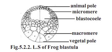
At the final stages of cleavage the embryo acquires a characteristic, mild, oblong shape. In this stage it is called the morula. The morula initially contains a shallow cavity called the blastocoele. Gradually the blastocoelic space increases into a large cavity occupying the middle of the blastula. However the blastocoele mostly remains in the animal pole region in the middle of the micromeres.
The blastomeres gradually adhere to each other, and arrange themselves into a true epithelium called the blastoderm. The blastoderm remains two cells thick in the animal pole. The embryo having a fluid-filled blastocoele and blastoderm is called the blastula.
It has been reported that around 12th cleavage the blastula possesses about 4096 cells. The blastula moves to the next stage, namely gastrulation at a stage in which it has about 20,000 cells.
The ultimate blastula is a ball of blastomeres which have to form different embryonic body layers and organs of the body. The fate of each and every blastomere has been observed and marked. A map showing various organ forming, areas on the blastula is called the ‘fate map’. This map shows prospective ectoderm, mesoderm and endodermal areas. It also shows the ‘zone of involution’ and ‘zone of invagination’ for the next stage of gastrulation.
Gastrulation in frog embryo
The process of gastrulation is a continuous activity succeeding, cleavage. During this process the blast-dermal cells begin to move. They wander and occupy their prospective organ forming zones. During this movement at one region on the blastula, the cells wander inside and occupy the blastocoelic cavity.
At a specific region below the equator the blastoderm cells assume an elongated bottle like shape. They move toward the interior of the blastula. As the cells move further inside, an invagination happens. A deepening of the invagination results in a cavity called the archenteron or gastrocoel.The opening of the archenteron on the surface of the blastula is called the blastopore.
The blastopore gradually assumes a crescentic shape. Finally it becomes circular. The region dorsal to the blastoporal opening is called the ‘dorsal lip'. The lower edge maybe called ventral lip

The surface cells representing several prospective zones of the embryo begin to wander inside through the blastopore. These inwandering of cell is termed as involution
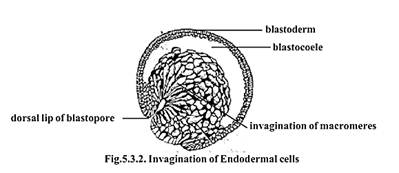
Initially, the first pharyngeal endodermal cells undergo invagination over the dorsal blastoporal lip. These cells move to the interior. They are followed by other cells. The inwandering cells gradually occupy the region of the blastocoele. Thus the blastocoelic cavity gets reduced. A new cavity among the involuted cells results. It is called the gastrocoel. The gastrocoel later becomes the archenteron. The interior region of the archenteron gradually transforms into the pharyngeal region. This region remains as the foregut. The mesodermal and endodermal cells gradually occupy their positions.
The inward movement of the exterior cells through the blastoporal region is called involution. The involution results in the positioning of chor-damesodermal cells and pharyngeal endodermal cells.
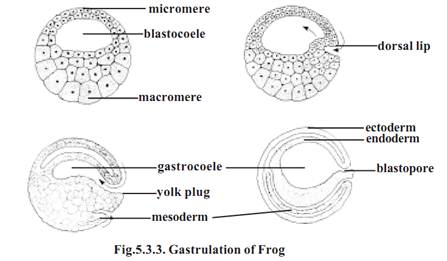
The mesodermal cells occupy the region between inner endodermal and outer ectodermal cells. While the exterior chorda-mesodermal cell involute inside, their place is taken up by the ectoderm. The expansion of the ectoderm is due to epiboly. Epiboly causes overlapping or ‘the roofing over’ of the gastrula by the ectoderm.
The blastopore is gradually covered by certain endoderm cells. The closing cells of the blastopore constitute the yolk-plug. Gradually the yolk-plug withdraws to the interior and the blastopore gets reduced into a narrow slit.
The process of gastrulation converts the blastula into a spherical, bilaterally symmetrical, triploblastic gastrula. Gradually the gastrula undergoes the process of tubulation or neurulation to become a neurula.
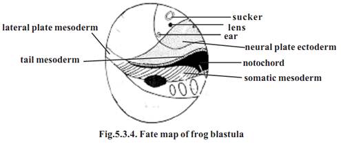
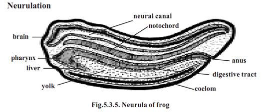
The process of neurulation is the formation of a neural tube. However during this process mesoderm and endoderm also undergo differentiation.
During neurulation the embryo lengthens along the anteroposterior axis. The dorsal side of the gastrula is lined by ectodermal cells. The presumptive area of the nervous system gets differentiated from the rest of ectoderm. It remains as medullary plate or nerual plate. The neural plate later thickens and it gets raised above the general level as ridges called neural folds. In the middle of the neural fold a neural groove appears. The neural groove deepens inside. The neural folds above the groove. The neural groove gets converted into a neural tube. This tube gets detached from the surface. The neural tube remains as the prospective nervous system. The embryo at this stage is called the neurula.
During neurulation, the tubulation of chorda-mesoderm and tubulation of endoderm also happen.
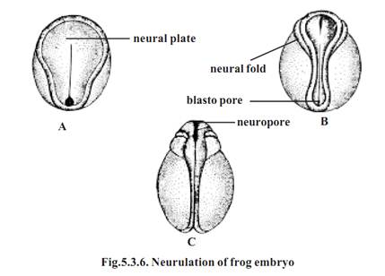
The postneurular development of frog involves the formation of all body organs.
Organogenesis of Frog
The primary organ rudiments from ectoderm, mesoderm and endoderm get well established during the processes of gastrulation and neurulation. In the next stage the primary organ rudiments subdivide into secondary organ rudiments. These rudiments get differentiated into various organs and organ systems.
The development of ectodermal organs
The neurula of frog has three kinds of ectodermal tissues namely, epidermal ectoderm, neural ectoderm and neural crest cells.
Epidermal ectoderm
The epidermal derivatives are the skin, olfactory sense organs, ear, lateral line sense organs, median fins, external gills and lining of mouth and anus.
Neural ectoderm
This layer of cells forms the central nervous system and peripheral nervous systems.
The development of mesodermal organs
The mesodermal derivatives are the limbs, endoskeleton, heart, blood vessels, kidney, coelom and reproductive organs.
The development of endodermal organs
The predominant endodermal organs are the organs of the alimentary canal, lungs, pancreas and urinary bladder.
Development of heart in Frog
The heart is a mesodermal derivative. It develops on the ventral side of pharynx. It is formed from the lateral plate mesoderm. Initially the heart is formed as a straight tube. Later it gets folded to form the chambered heart.
References
T. Sargunam Stephen, Biology (Zoology). First Edition – 2005, Government of Tamilnadu.


|
|
|
|
تفوقت في الاختبار على الجميع.. فاكهة "خارقة" في عالم التغذية
|
|
|
|
|
|
|
أمين عام أوبك: النفط الخام والغاز الطبيعي "هبة من الله"
|
|
|
|
|
|
|
قسم شؤون المعارف ينظم دورة عن آليات عمل الفهارس الفنية للموسوعات والكتب لملاكاته
|
|
|