

 النبات
 الحيوان
 الأحياء المجهرية
 علم الأمراض
 التقانة الإحيائية
 التقنية الحيوية المكروبية
 التقنية الحياتية النانوية
 علم الأجنة
 الأحياء الجزيئي
 علم وظائف الأعضاء
 الغدد
الغدد
 المضادات الحيوية
|
Read More
Date: 20-7-2021
Date: 25-2-2021
Date: 4-1-2021
|
Osteology
Bones of the lower limb (proximal to distal) include the hip (os coxae), femur, patella, tibia, fibula, tarsal bones, metatarsals, and phalanges.
A. Hip
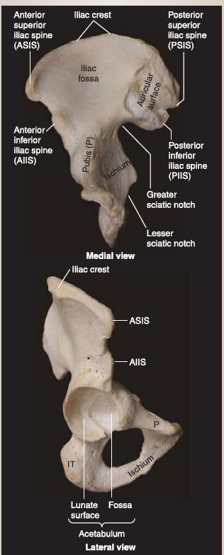
As shown in Figure 1, in the adult, each hip bone is formed by the fusion of three bones: ilium, ischium, and pubis. The acetabulum, which serves as the socket portion of the hip joint, is formed where all three bones intersect laterally. Right and left hip bones are joined posteriorly by the sacrum through the sacroiliac joint and anteriorly at the pubis symphysis-forming the ring-like pelvic girdle.


Figure 1: Osteology of the hip bone.
B. Femur
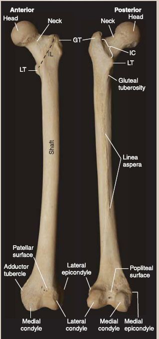
The femur (thigh bone) is the longest and heaviest bone in the body (Fig. 2). Proximally, the femur is characterized by a round femoral head (ball portion of the hip joint) and inferolaterally angled neck. The neck connects the head to the shaft of the femur adjacent to the greater and lesser trochanters-two bony prominences that serve as muscle and ligament attachment sites. The trochanters are connected posteriorly by the intertrochanteric crest and anteriorly by the intertrochanteric line. The femoral shaft is primarily smooth, except for the raised ridge of bone posteriorly, the linea aspera. Distally, the femur has rounded medial and lateral condyles that articulate with
the proximal tibia to form the knee joint. The condyles are separated by an intercondylar fossa posteriorly and inferiorly. Medial and lateral epicondyles extend superiorly from the condyles and serve as collateral ligament attachment sites. An adductor tubercle is located just superior to the medial epicondyle of the femur and serves as a tendon attachment site.

Figure 2: Osteology of the femur. GT = greater trochanter, IC = intertrochanteric crest, LT = lesser trochanter.
C. Patella
The patella (knee cap) is a triangular-shaped sesamoid bone that covers the anterior intercondylar surface of the femur and contributes to the kinetics of the knee joint.
D. Tibia
The tibia (shin bone) is the weight-bearing bone of the leg (Fig. 3). Proximally, it is characterized by flat, concave lateral and medial tibial condyles that articulate with the distal femur. Between the tibial condyles are the intercondylar eminence and tubercles,
which serve as ligament attachment sites. Anteriorly, the tibial tuberosity is a palpable mass onto which the patellar ligament attaches. Posterolaterally, there is a concave facet where the fibular head articulates. The shaft of the tibia is marked by a lateral interosseous border and sharp anterior ridge that extends distally, where the bone tapers to articulate with the talus and distal fibula. The medial malleolus is the distal most part of the tibia and frames the ankle joint medially.

Figure 3: Osteology of the tibia
E. Fibula
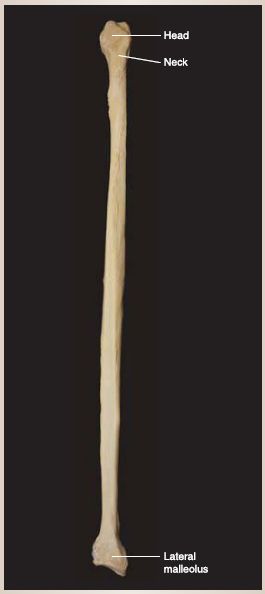
The fibula is a thin, non-weight-bearing bone in the leg that articulates laterally with the tibia. Proximally, it is characterized by an arrowheadshaped head and narrow neck, which are easily palpated superficially (Fig. 4). The interosseous border lines the medial shaft. Distally, the fibula expands into the lateral malleolus, which frames the ankle joint laterally.

Figure 4: Osteology of the fibula.
F. Tarsal bones
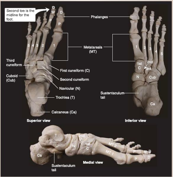
The tarsal bones include the talus, calcaneus, cuboid, navicular, and three cuneiforms (1-3), as shown in Figure 5. Of the seven tarsal bones, only the talus articulates with the tibia and fibula to form the ankle joint. Subtalar tarsal bones contribute to the shape and function of the foot. The talus is characterized by a dome-shaped trochlea, which is framed by the medial and lateral malleoli. The calcaneus (heel bone) articulates with the body of the talus inferiorly and with the cuboid anteriorly. It is the largest and strongest talar bone in the foot. The calcaneus has a shelf-like projection laterally, called the sustentaculum tali, which supports the talus, and a posteroinferior calcaneal tuberosity, which bears body weight in the hind foot. Medially, the boat-shaped navicular bone lies between the talus and three cuneiforms, while the cuboid lies laterally to complete the distal tarsal row.

Figure 5 : Osteology of the foot. D = distal, M = middle, P = proximal.
G. Metatarsals and phalanges
Five metatarsal bones (1-5; medial to lateral) contribute to the forefoot and articulate with the distal row of tarsal bones (cuneiforms and cuboid). Each metatarsal has a proximal base, middle shaft, and distal head. The head of each metatarsal articulates with the base of the proximal phalange. The first digit (hallux; big toe) has two phalanges-proximal and distal-while the remaining four lateral digits have three-proximal, middle, and distal.
H. Surface anatomy
Several surface landmarks of the lower limb are important during physical examination . Assessing symmetry of the kinetic chain, from the pelvis down to the feet, is essential when formulating a diagnosis. Symmetry and level of these landmarks can be assessed in sitting, standing, or supine positions to determine where along the kinetic chain an underlying deviation may be causing impairment.
1. Bony landmarks: In the pelvis, these include the anterior and posterior superior iliac spines (S2 level), iliac crests (L4 level), pubic symphysis, and ischial tuberosities. Femoral bony landmarks include the greater trochanters and condyles. Landmarks in the leg include the tibial tuberosity, fibular head, medial malleolus, and lateral malleolus. At the ankle, the talus can be palpated between the medial and lateral malleoli.
2. Soft tissue landmarks: These include the gluteal, inguinal, and popliteal creases.


|
|
|
|
علامات بسيطة في جسدك قد تنذر بمرض "قاتل"
|
|
|
|
|
|
|
أول صور ثلاثية الأبعاد للغدة الزعترية البشرية
|
|
|
|
|
|
|
مكتبة أمّ البنين النسويّة تصدر العدد 212 من مجلّة رياض الزهراء (عليها السلام)
|
|
|