

 النبات
 الحيوان
 الأحياء المجهرية
 علم الأمراض
 التقانة الإحيائية
 التقنية الحيوية المكروبية
 التقنية الحياتية النانوية
 علم الأجنة
 الأحياء الجزيئي
 علم وظائف الأعضاء
 الغدد
الغدد
 المضادات الحيوية
|
Read More
Date: 9-1-2017
Date: 6-8-2016
Date: 2-8-2016
|
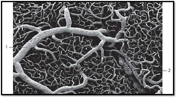
Capillary Network-Lacrimal Gland
The capillary bed of an organ can be made visible using one of several injection techniques, and its three-dimensional structure can then be examined by scanning electron microscopy. In this instance, the arteries that go to the head of a cat were filled with a resin, which was allowed to harden at a defined temperature range. Organic materials were than removed using acid or alkaline solutions (maceration ). This procedure creates a corrosion preparation, which leaves the geometry of the capillaries intact. This figure shows the dense capillary network in the lacrimal gland of a cat. Note the branching of the larger arteries 1 . Veins 2 are visible in the bottom left of the figure. Differences in form and orientation of the endothelial cell nuclei, among other attributes, indicate whether a vessel is an artery or vein.
1 Arteries
2 Veins
Scanning electron microscopy; magnification: × 85

References
Kuehnel, W.(2003). Color Atlas of Cytology, Histology, and Microscopic Anatomy. 4th edition . Institute of Anatomy Universitätzu Luebeck Luebeck, Germany . Thieme Stuttgart · New York .


|
|
|
|
علامات بسيطة في جسدك قد تنذر بمرض "قاتل"
|
|
|
|
|
|
|
أول صور ثلاثية الأبعاد للغدة الزعترية البشرية
|
|
|
|
|
|
|
مكتبة أمّ البنين النسويّة تصدر العدد 212 من مجلّة رياض الزهراء (عليها السلام)
|
|
|