

 النبات
 الحيوان
 الأحياء المجهرية
 علم الأمراض
 التقانة الإحيائية
 التقنية الحيوية المكروبية
 التقنية الحياتية النانوية
 علم الأجنة
 الأحياء الجزيئي
 علم وظائف الأعضاء
 الغدد
الغدد
 المضادات الحيوية
|
Read More
Date: 8-1-2017
Date: 15-1-2017
Date: 2-8-2016
|
Kinocilia-Uterine Tube
The detail structure of cilia is particularly impressive in cross-sections: there are two tubules in the center, and nine pairs of tubules (doublets) are located around them in the periphery (9 × 2 + 2 pattern). The peripheral twin tubules ( microtubule doublets, axoneme) are close to each other and share a common central wall. The wall consists of two or three protofilaments that are pre-dominantly compose d of the protein tubulin (α- and β-tubulin). This particular arrangement creates two different tubules within the twin tubules (doublet); only one of them, the A -microtubule , features a complete ring structure with 13 protofilaments. The B -tubule forms an incomplete ring. The B- tubule itself consists of only nine protofilaments. Three or four protofilaments in its structure belong to the A-ring. In contrast to the central microtubule pair, the A-tubules at the periphery have small “arms,” which reach to the adjacent pairs of microtubules. This figure shows these arms only vaguely. The arms consist of the protein dynein and ATPase. They play an important role in the motility of cilia. The two central microtubules are separate, side-by-side units. The peripheral axoneme are clearly connected to each other via the binding protein (nexin ). Axial view of the ciliate d cells from oviduct.
Electron microscopy; magnification: × 60 000

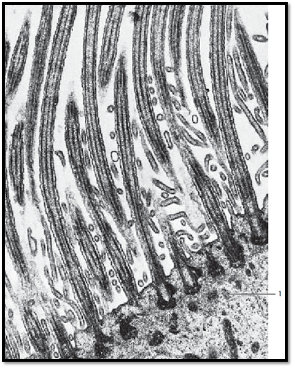
Longitudinal section through the kinocilia of a ciliate d epithelial cell from oviduct. Between the cilia are several sections through arcuate microvilli in different angles. Inside the cilia, microtubules form the longitudinal axis (axoneme), which consist of two central tubules (central tubule pair) and nine peripheral double tubules (doublets, each made of A - and B-tubules): 9 × 2 + 2 structure. The nine parallel outer axial tubules are connected to the central two tubules via spoke proteins. The two peripheral double tubules are different: in an axial section, tubule A forms a ring. In contrast, tubule B look s like an incomplete circle, like the letter C. The peripheral ciliary microtubules extend into the apical cytoplasm region. There, underneath the cell surface, they form basal bodies ( basal nodes, kinetosomes, cilia root ) . Kinetosomes form the cytoplasmic anchor places for microtubules. Their structure is that of centrioles. At the basal side, there is often a structure called rootstock , which shows a striation pattern. The rootstock contains the protein centrin . Kinetosomes are densely packed in richly ciliate d epithelium. This accounts for the layer of basal bodies ( basal nodule row ) that is visible in light microscopy.
1 Apical cytoplasm in a ciliated cell
Electron microscopy; magnification: × 30 000

References
Kuehnel, W.(2003). Color Atlas of Cytology, Histology, and Microscopic Anatomy. 4th edition . Institute of Anatomy Universitätzu Luebeck Luebeck, Germany . Thieme Stuttgar t · New York .


|
|
|
|
دخلت غرفة فنسيت ماذا تريد من داخلها.. خبير يفسر الحالة
|
|
|
|
|
|
|
ثورة طبية.. ابتكار أصغر جهاز لتنظيم ضربات القلب في العالم
|
|
|
|
|
|
|
العتبة العباسية المقدسة تستعد لإطلاق الحفل المركزي لتخرج طلبة الجامعات العراقية
|
|
|