

 النبات
 الحيوان
 الأحياء المجهرية
 علم الأمراض
 التقانة الإحيائية
 التقنية الحيوية المكروبية
 التقنية الحياتية النانوية
 علم الأجنة
 الأحياء الجزيئي
 علم وظائف الأعضاء
 الغدد
الغدد
 المضادات الحيوية
|
Read More
Date: 10-7-2021
Date: 2025-02-05
Date: 9-7-2021
|
Pelvic Muscles and Fascia
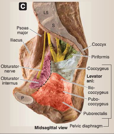
Muscles of the pelvic wall assist in supporting pelvic viscera by partially closing off the pelvic outlet (Fig. 1). These muscles include obturator internus, piriformis, coccygeus, and levator ani, with the latter two forming the pelvic diaphragm. Obturator internus and piriformis externally rotate the thigh a.Pelvic fascia provides additional support and protection to pelvic viscera.


Figure 1: Pelvic floor muscles. A and B. Pelvic musculature with organs removed. C. Cadaveric specimen with pelvic viscera and vasculature removed. Dashed black line represents tendinous arch. A = anal canal site, LST = lumbosacral trunk, P = pubic symphysis, S = sacrum, U = urethra site, V = vagina site.
A. Pelvic diaphragm
Coccygeus and levator ani collectively form the pelvic diaphragm, which serves as the floor of the pelvic cavity. This hammock-like muscular diaphragm is reinforced by pelvic fascia and interrupted in the anterior midline at the urogenital hiatus. The urogenital hiatus allows for the passage of the urethra and vagina in females and the intermediate (membranous) urethra in males. Pelvic diaphragm muscles are innervated by the nerve to levator ani and coccygeus (S3-S4).
1. Coccygeus: Located inferior to the piriformis internally, coccygeus originates from the ischial spine and inserts onto the sacrum and coccyx. It functions to raise the pelvic floor by flexing the coccyx anteriorly.
2. Levator ani: Levator ani is made up of three muscles, which collectively form the majority of the pelvic diaphragm. Together, these muscles raise the pelvic floor, which increases intra-abdominal pressure during urination, defecation, vomiting, and parturition.
a. Pubococcygeus: Originating anteriorly on the pubis and inserting onto the coccyx, pubococcygeus can be further differentiated by its anterior fibers that form a supportive sling around the vagina (pubovaginalis) or prostate (puboprostaticus). These fibers insert directly into, and further strengthen, the perinea! body.
b. Puborectalis: Located inferomedially to pubococcygeus and also originating from the pubis, this muscle forms a U-shaped sling (rectal sling) at the level of the anorectal junction. In addition to supporting the pelvic floor, it functions to pull the anorectal junction anteriorly to support fecal mass. Relaxation of puborectalis allows for fecal mass to pass into the anal canal for defecation.
c. lliococcygeus: lliococcygeus originates from the ischial spine and tendinous arch and inserts into the coccyx.
B. Pelvic fascia
Pelvic fascia is a connective tissue matrix that fills or lines spaces around viscera and between the parietal peritoneum of the abdominal cavity and the muscular walls and floor of the pelvic cavity. It is described as having visceral and parietal (membranous) components, which are separated by endopelvic fascia.
1. Visceral pelvic fascia: This layer lines the pelvic viscera and is continuous with parietal fascia where the organs pass through the pelvic hiatuses-urogenital and rectal.
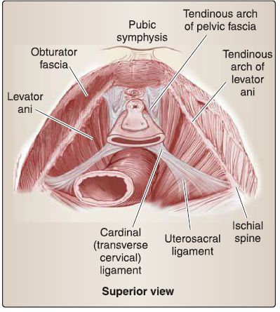
2. Parietal pelvic fascia: This layer lines the muscles of the pelvic walls and floor internally. Selective areas are thicker than others, such as the obturator fascia and tendinous arch of levator ani.
3. Endopelvic fascia: Endopelvic fascia occupies spaces around pelvic viscera (Fig. 2). Described as either loose areolar connective tissue or dense fibrous tissue-often referred to as ligaments-this fascia functions to support and protect pelvic viscera, while allowing for distension of viscera. Two of the most prominent ligaments formed by endopelvic fascia are the paired transverse cervical (cardinal} and uterosacral ligaments in the female pelvis. Additional supportive structures include the lateral ligaments of the bladder and rectum.
a. Transverse cervical (cardinal} ligament: Located at the broad ligament base, these ligaments support the cervix by connecting
it to the pelvic walls laterally and transmit uterine arteries and
veins.
b. Uterosacral: These ligaments help to maintain the position of
the uterus by connecting the cervix to the sacrum.

Figure 2 : Pelvic fascia.


|
|
|
|
لخفض ضغط الدم.. دراسة تحدد "تمارين مهمة"
|
|
|
|
|
|
|
طال انتظارها.. ميزة جديدة من "واتساب" تعزز الخصوصية
|
|
|
|
|
|
|
بمناسبة مرور 40 يومًا على رحيله الهيأة العليا لإحياء التراث تعقد ندوة ثقافية لاستذكار العلامة المحقق السيد محمد رضا الجلالي
|
|
|