

 النبات
 الحيوان
 الأحياء المجهرية
 علم الأمراض
 التقانة الإحيائية
 التقنية الحيوية المكروبية
 التقنية الحياتية النانوية
 علم الأجنة
 الأحياء الجزيئي
 علم وظائف الأعضاء
 الغدد
الغدد
 المضادات الحيوية
|
Read More
Date: 28-7-2016
Date: 9-1-2017
Date: 23-1-2017
|
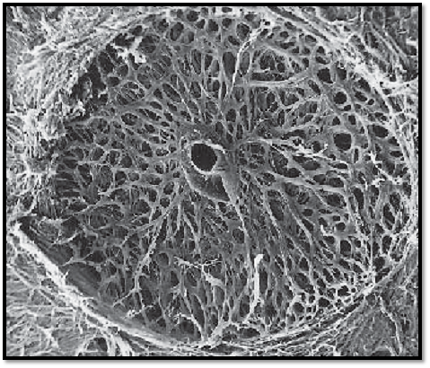
Optic Nerve-Lamina Cribrosa Sclerae
The optic nerve, a longitudinal fascicle, has an intraocular, orbital, intracanalicular and an intracranial segment. The about 2-mm long pars intraocularis corresponds to the papilla of the optic nerve. The nerve f ibers in this intraocular segment are unmyelinated. Examination with the ophthal-moscope allows a view of the sieve-like, loosely structured lamina cribrosa sclerae, which shines through the unmyelinated nerve fibers at the point where the optic nerve traverses the lamina cribrosa. The lamina cribrosa is a continuation of the sclera. The axons of the optic nerve are myelinated only after traversing the lamina cribrosa. This figure shows the lamina cribrosa sclerae. The viewer look s on the circular arrangements of collagen fibers around each traversing axon. The collagen f ibers have been isolate d by maceration with a 10% NaOH solution, which disintegrates the axons of the optic nerve. The center represents the space for the artery and the central retinal vein. The collagen fibers around the center are called scleral ring . Lamina cribrosa sclerae of an 89-year-old woman.
Scanning electron microscopy; magnification: × 60

References
Kuehnel, W.(2003). Color Atlas of Cytology, Histology, and Microscopic Anatomy. 4th edition . Institute of Anatomy Universitätzu Luebeck Luebeck, Germany . Thieme Stuttgart · New York .


|
|
|
|
التوتر والسرطان.. علماء يحذرون من "صلة خطيرة"
|
|
|
|
|
|
|
مرآة السيارة: مدى دقة عكسها للصورة الصحيحة
|
|
|
|
|
|
|
نحو شراكة وطنية متكاملة.. الأمين العام للعتبة الحسينية يبحث مع وكيل وزارة الخارجية آفاق التعاون المؤسسي
|
|
|