


 النبات
 الحيوان
 الأحياء المجهرية
 علم الأمراض
 التقانة الإحيائية
 التقنية الحيوية المكروبية
 التقنية الحياتية النانوية
 علم الأجنة
 الأحياء الجزيئي
 علم وظائف الأعضاء
 الغدد
الغدد
 المضادات الحيوية
|
Read More
Date: 4-11-2015
Date: 4-11-2015
Date: 4-11-2015
|
Earthworm
Phylum - Annelida
Class - Chaetopoda
Order - Oligochaeta
Type - Lampito mauritii
Earthworms are nocturnal animals. They lie in the burrows during the day and come out at night for food. Earthworms leave the burrow only during the rainy season when their burrows are flooded with water.
External features
Lampito (Megascolex) mauritii is a common earthworm found in South India. The body is long, slender, cylindrical and bilaterally symmetrical. It is about 8 to 21 cm long and 3 to 4 mm in thickness. The dorsal surface is dark purplish brown, and the ventral surface is paler in colour. It is marked by a series of segments. The segments are separated from one another by inter- segmental grooves. The division is both external and internal. Inside the body, each cavity of the segment is separated from the next, by a thin partition called the septum. All the segments look alike. This kind of repetitive arrangement of the segments is called metamerism.
The mouth is found in the center of the first segment of the body, called the peristomium. Overhanging the mouth is a small flap called the upper lip or prostomium. The last segment has the anus. It is called the pygidium. In mature worms, segments 14 to 17 may be found swollen with a glandular thickening of the skin called clitellum.
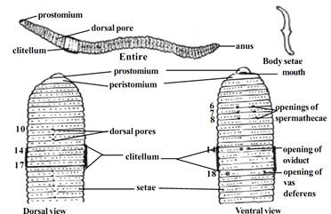
Fig. 1.3.2. Earthworm-External
Body setae
Tiny curved bristles called setae are found embedded in small pits of the body wall. These pits are called the setigerous pits. The setae are arranged around the body. They are made of chitin and have a swollen middle part and pointed curved ends. The setae resemble the mathematical symbol .They can be moved in any direction and extended or withdrawn by the action of muscles. They are used for locomotion
External apertures:
i) Dorsal pores: These are minute openings situated in the mid dorsal line in the intersegmental grooves commencing from the 10th segment. The coelom communicates to the exterior through these pores and keep the body surface moist and free from harmful micro organisms.
ii) Spermathecal openings: Three pairs of openings are situated ventrolaterally in the intersegmental grooves between segments six and seven, seven and eight and eight and nine. These opening can be easily seen in mature worms.
iii) Openings of oviduct: These are a pair of apertures lying close together on the ventral surface of the 14th segment.
iv) Openings of Spermiduct: A pair of apertures is situated on the lowerside of the 18th segment.
v) Nephridiopores: Numerous minute openings scattered on the body wall from 14th segment onwards.
Body wall:
The body wall of earthworm is thin soft and moist. It consists of the following layers arranged from outside.
Cuticle: It is a thin, transparent, iridescent layer secreted by the underlying epidermis.
Epidermis: It is in the form of a single layer of columnar cells. This layer contains gland cells and receptor cells.
Dermis: It is a very thin sheet of connective tissue forming a basement for the epithelial cells on the outside and muscles on the inside.
Muscles: The muscles are arranged in two layers, namely the outer circular and inner longitudinal.
Coelomic epithelium: It is the inner most layer of the body wall forming the lining of the body cavity.
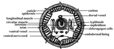
Fig. 1.3.3. Earthworm-T.S.
Body Cavity:
A spacious body cavity called the coelom is seen between the alimentary canal and the body wall. It is divided into a series of compartments by the transverse partitions of connective tissue called the septa. The coelom is lined with the coelomic epithelium and filled with coelomic fluid. It is a colorless fluid with amoeboid coelomic corpuscles floating in it. The fluid oozes out through the dorsal pores. It keeps body surface moist as a condition quite essential for respiration. The coelomic cavity communicates to the exterior through reproductive and excretory apertures. The germ cells are budded off from the wall of this cavity.
Locomotion:
Earthworms move about by contraction and expansion of its body wall. When the circular muscles of the body wall contract, the body becomes thin and elongated. This process results in the forward extension of the body. Then it fixes itself firmly to the ground with help of the body setae and mouth. Subsequently when the longitudinal muscles contract, the body becomes thick and shortened. As a result, the body is drawn forward towards the anterior end which is already fixed to the ground. Thus by a repeated process of alternate contraction and expansion of muscular body wall locomotion is effected.
Digestive System:
The digestive system runs as a straight tube from mouth to anus. The mouth is situated in the first segment. The mouth opens into the buccal cavity which occupies segments 1 and 2. The buccal cavity in turn leads into a thick muscular Pharynx. The pharynx occupies segments 3 and 4 and is surrounded by the pharyngeal glands. The oesophagus is a short narrow tube lying in 5th segment. It leads into the gizzard lying in the 6th segment. Its inner surface has a chitinous lining. The intestine is a large tube extending from the gizzard to the anus. The intestine up to the 14th segment is narrow and the remaining part is sacculated. The dorsal wall of the intestine is folded into the cavity as the typhlosole. This fold contains blood vessels. It increases the absorptive area of the intestine. The inner epithelium consists of columnar cells and glandular cells.
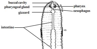
Fig. 1.3.4. Earthworm-Digestive system
Feeding:
The earthworm feeds on decaying organic materials contained in the soil. It takes the soil into its alimentary canal where the organic matter is digested and absorbed. The unwanted soil particles are sent out as worm casts.
Circulatory System:
In the body of earthworm there are two median longitudinal vessels. The dorsal longitudinal vessel runs above the alimentary canal. The ventral longitudinal vessel runs below the alimentary canal. The dorsal vessel is contractile and blood flows forwards in it. There are paired valves inside this vessel which prevent the backward flow of the blood. The ventral vessel is non-contractile and blood flows backwards in it. The ventral vessel has no valves. In the anterior part of the body the dorsal vessel is connected with the ventral vessel by eight pairs of commissural vessels or the lateral hearts lying in the segments 6 to 13. These vessels run on either side of the alimentary canal and pump blood from the dorsal vessel to the ventral vessel.
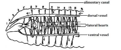
Fig. 1.3.5. Earthworm - Circulatory system-Lateral view
The dorsal vessel receives blood from various organs in the body. The ventral vessel supplies blood to the various organs.
Excretory System:
Excretion is the process of elimination of metabolic waste products from the body. In earthworm, excretion is effected by minute paired tubes called nephridia. These are found, one pair, in each segment.
A typical nephridium has an internal funnel like opening called the nephrostome. It is fully ciliated. The nephrostome is in one segment and the rest of the tube will be in the succeeding segment. This tube has three distinct
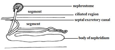
Fig. 1.3.6. Earthworm - nephridium
divisions. The first part following the nephrostome is ciliated inside. This is called the ciliated region. The next part is wider and is thrown into coils. This part has glands on its wall. It is called the glandular region. The last part has neither cilia nor glands. It is called the muscular region. This region opens outside by an aperture called the nephridiopore. The waste material is collected from the body cavity by the ciliated funnel. The ciliated region pushes the waste into the nephridium. The glandular part extracts waste from the blood and add it on to the waste inside. Finally the waste goes out through the nephridiopore.
In the South Indian earthworm, Megascolex, there are certain modifications. There are three types of nephridia in the megascolex. They are: (i). Meganephridia, (ii). Micronephridia, (iii). Pharyngeal nephridia.
Besides nephridia there are some special cells on the wall of the intestine called Chlorogogen cells. They collect the waste and then drop down into the body cavity. These are then sent out through nephridia.
Nervous System:
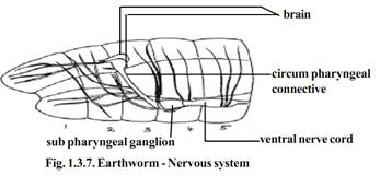
The brain is formed of the supra pharyngeal ganglia. It is a bilobed mass of nervous tissue situated on the dorsal wall of the pharynx in 3rd segment. The ganglia found below the pharynx in the 4th segments is called the subpharyngeal ganglia. The brain and the subpharyngeal ganglia are connected by a pair of circum pharyngeal connectives. They run one on each side of the pharynx. Thus a nerve ring is formed around the anterior region of the alimentary canal. The double, solid ventral nerve cord runs backwards from the subpharyngeal ganglia, in the mid ventral line to the hind end of the body. The ventral nerve cord has segmental ganglion one in each segment. From the brain nerves are given off to the peristomium. From each ganglion of the ventral nerve cord, three pairs of nerves are given off to the body wall and other organs.
Receptors which are stimulated by the sense of touch (tactile receptors), Chemical changes (chemoreceptors) and changes in temperature (thermoreceptors) are present in the body wall. These receptors are in the form of groups of slender columnar cells with short hairs projecting at the free end and connected with sensory fibers at the inner end. Receptors stimulated by changes in the intensity of light (Photoreceptors) are found on the dorsal surface of the body. Gustatory receptors (sense of taste) and olfactory receptors (sense of smell) are found in the buccal cavity.
Reproductive System:
Both male and female reproductive organs are present in the same worm. Hence the earthworms are known as hermaphrodites. Since the sperms develop earlier than production of ova, self-fertilization is avoided. It is known as protandry.
Male reproductive organs:
The male sex organs are formed of two pairs of testes and a pair of vasa deferentia. Testes are found in segments 10 and 11. They are tufts of finger shaped processes attached to the anterior septa of segments 10 and 11. There are two pairs of seminal versicles formed as outgrowths of the testicular segments. Further two pairs of seminal funnels called ciliary rosettes are situated in the same segment as the testes. The ciliated funnels of the same side are connected to a long tube called vas deferens. The two vasa deferentia of both sides run backwards along the ventral body wall up to the 18th segment where they open to the exterior through the male genital aperture. Male genital apertures contain penial setae for copulation. A pair of prostate glands, each in the form of a much coiled tube are situated in segments 18 and 19. The prostate glands open to the exterior along with the vas deferens. The secretion of the prostate glands help to arrange the sperms into bundles called spermatophores.
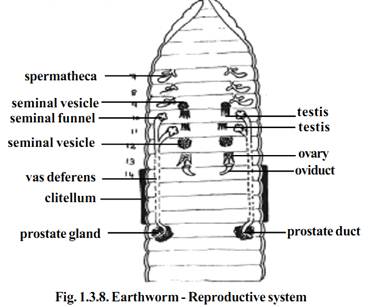
Female reproductive organs.
Pair of ovaries is found lying in segment 13. They are attached to the anterior septum of the 13th segment. Each ovary is a flat structure with a number of fingers like processes. The ova are arranged in a linear order in the ovaries. There is pair of oviducts. They open internally into the 13th segment and externally on the ventral surface of the 14th segment. Three pairs of spermathecae are present in segments 7, 8 and 9. These external openings are situated in the intersegmental grooves of segments 6 and 7, 7 and 8, and 8 and 9. The spermatozoa received from another individual during copulation are stored in spermathecae.
Copulation:
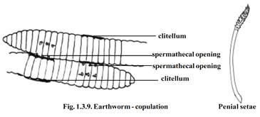
During copulation the head ends of the two worms are directed in the opposite directions and the clitellum of one worm is opposite to the Spermathecal segments of the other. The spermatozoa of one worm pass into the spermathecae of the other worms. The worms separate after the mutual exchange of spermatozoa.
Later the glandular cells of the clitellum secrete a thick fluid which hardens into a girdle surrounding the clitellum
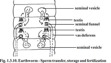
The girdle is moved forward by the wriggling movements of the body. As the girdle is moved forwards it receives the ova and spermatozoa. The girdle containing the germ cells (ova and sperms) and the nutrient albuminous fluid is slipped off at the anterior end and it becomes a closed sac called the cocoon. Fertilization and the development of the eggs into worms take place within the cocoon. Young worms come out of the cocoon after complete development.
References
T. Sargunam Stephen, Biology (Zoology). First Edition – 2005, © Government of Tamilnadu.


|
|
|
|
للعاملين في الليل.. حيلة صحية تجنبكم خطر هذا النوع من العمل
|
|
|
|
|
|
|
"ناسا" تحتفي برائد الفضاء السوفياتي يوري غاغارين
|
|
|
|
|
|
|
شعبة فاطمة بنت أسد (عليها السلام) تنظم دورة تخصصية في أحكام التلاوة
|
|
|