
النبات

مواضيع عامة في علم النبات

الجذور - السيقان - الأوراق

النباتات الوعائية واللاوعائية

البذور (مغطاة البذور - عاريات البذور)

الطحالب

النباتات الطبية


الحيوان

مواضيع عامة في علم الحيوان

علم التشريح

التنوع الإحيائي

البايلوجيا الخلوية


الأحياء المجهرية

البكتيريا

الفطريات

الطفيليات

الفايروسات


علم الأمراض

الاورام

الامراض الوراثية

الامراض المناعية

الامراض المدارية

اضطرابات الدورة الدموية

مواضيع عامة في علم الامراض

الحشرات


التقانة الإحيائية

مواضيع عامة في التقانة الإحيائية


التقنية الحيوية المكروبية

التقنية الحيوية والميكروبات

الفعاليات الحيوية

وراثة الاحياء المجهرية

تصنيف الاحياء المجهرية

الاحياء المجهرية في الطبيعة

أيض الاجهاد

التقنية الحيوية والبيئة

التقنية الحيوية والطب

التقنية الحيوية والزراعة

التقنية الحيوية والصناعة

التقنية الحيوية والطاقة

البحار والطحالب الصغيرة

عزل البروتين

هندسة الجينات


التقنية الحياتية النانوية

مفاهيم التقنية الحيوية النانوية

التراكيب النانوية والمجاهر المستخدمة في رؤيتها

تصنيع وتخليق المواد النانوية

تطبيقات التقنية النانوية والحيوية النانوية

الرقائق والمتحسسات الحيوية

المصفوفات المجهرية وحاسوب الدنا

اللقاحات

البيئة والتلوث


علم الأجنة

اعضاء التكاثر وتشكل الاعراس

الاخصاب

التشطر

العصيبة وتشكل الجسيدات

تشكل اللواحق الجنينية

تكون المعيدة وظهور الطبقات الجنينية

مقدمة لعلم الاجنة


الأحياء الجزيئي

مواضيع عامة في الاحياء الجزيئي


علم وظائف الأعضاء


الغدد

مواضيع عامة في الغدد

الغدد الصم و هرموناتها

الجسم تحت السريري

الغدة النخامية

الغدة الكظرية

الغدة التناسلية

الغدة الدرقية والجار الدرقية

الغدة البنكرياسية

الغدة الصنوبرية

مواضيع عامة في علم وظائف الاعضاء

الخلية الحيوانية

الجهاز العصبي

أعضاء الحس

الجهاز العضلي

السوائل الجسمية

الجهاز الدوري والليمف

الجهاز التنفسي

الجهاز الهضمي

الجهاز البولي


المضادات الميكروبية

مواضيع عامة في المضادات الميكروبية

مضادات البكتيريا

مضادات الفطريات

مضادات الطفيليات

مضادات الفايروسات

علم الخلية

الوراثة

الأحياء العامة

المناعة

التحليلات المرضية

الكيمياء الحيوية

مواضيع متنوعة أخرى

الانزيمات
Rickettsia, Coxiella, Orientia, and Ehrlichia
المؤلف:
Fritz H. Kayser
المصدر:
Medical Microbiology -2005
الجزء والصفحة:
8-3-2016
3206
Rickettsia, Coxiella, Orientia, and Ehrlichia
The genera of the Rickettsiaceae and Coxelliaceae contain short, coccoid, small rods that can only reproduce in host cells. With the exception of Coxiella (aerogenic transmission), they are transmitted to humans via the vectors lice, ticks, fleas, or mites. R. prowazekii and R. typhi cause typhus, a disease characterized by high fever and a spotty exanthem. Several rickettsiae species cause spotted fever, a milder typhus like disease. Orientia tsutsugamushi is transmitted by mite larvae to cause tsutsugamushi fever. This disease occurs only in Asia. Coxiella burnetii is responsible for Q fever, an infection characterized by a pneumonia with an atypical clinical course.
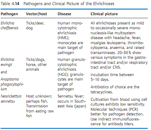
Several species of Ehrlichiaceae cause ehrlichiosis in animals and humans. The method of choice for laboratory diagnosis of the various rickettsioses and ehrlichioses is antibody assay by any of several methods, in most cases indirect immunofluorescence. Tetracyclines represent the antibiotic of choice for all of these infections. Typhus and spotted fever no longer occur in Europe. Q fever infections are reported from all over the world. Sources of infection include diseased sheep, goats, and cattle. The prognosis for the rare chronic form of Q fever (syn. Q fever endocarditis) is poor. Ehrlichiosis infects mainly animals, but in rare cases humans as well.
Classification. The bacteria of this group belong to the families Rickettsiaceae (Rickettsia and Orientia), Coxelliaceae (Coxiella), and Ehrlichiaceae (Ehrlichia, Anaplasma, Neorickettsia). Some of these organisms can cause mild, self- limiting infections in humans, others severe disease. Arthropods are the transmitting vectors in many cases.
Morphology and culture. These obligate cell parasites are coccoid, short rods measuring 0.3-0.5 µm that take gram staining weakly, but Giemsa staining well. They reproduce by intracellular, transverse fission only. They can be cultured in hen embryo yolk sacs, in suitable experimental animals (mouse, rat, guinea pig) or in cell cultures.
Pathogenesis and clinical pictures. With the exception of C. burnetii, the organisms are transmitted by arthropods. In most cases, the arthropods excrete them with their feces and ticks transmit them with their saliva while sucking blood. The organisms invade the host organism through skin injuries. C. burnetii is transmitted exclusively by inhalation of dust containing the pathogens. Once inside the body, rickettsiae reproduce mainly in the vascular endothelial cells. These cells then die, releasing increasing numbers of organisms into the bloodstream. Numerous inflammatory lesions are caused locally around the destroyed endothelia. Ehrlichiae reproduce in the monocytes or granulocytes of membrane-enclosed cytoplasmic vacuoles. The characteristic morulae clusters comprise several such vacuoles stuck together.
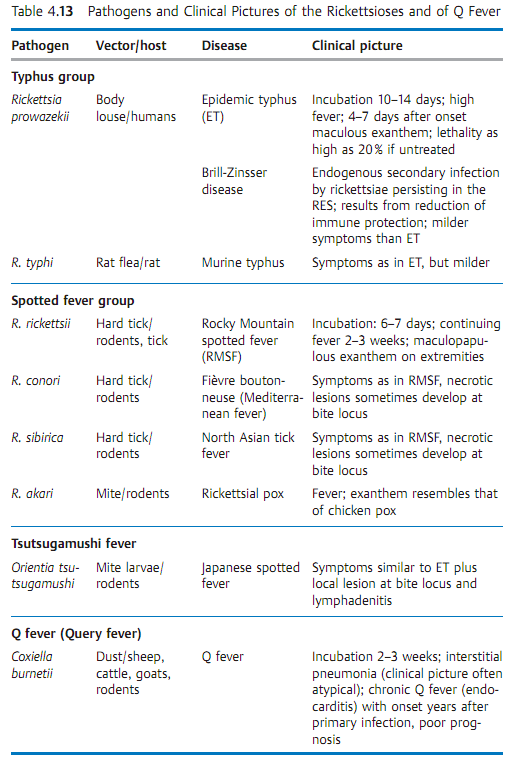
Table 4.13 summarizes a number of characteristics of the rickettsioses.
Diagnosis. Direct detection and identification of these organisms in cell cultures, embryonated hen eggs, or experimental animals is unreliable and is also not to be recommended due to the risk of laboratory infections. Special laboratories use the polymerase chain reaction to identify pathogen-specific DNA sequences. However, the method of choice is currently still the antibody assay, whereby the immunofluorescence test is considered the gold standard among the various methods. The Weil-Felix agglutination test is no longer used today due to low sensitivity and specificity.
Therapy. Tetracycline lower the fever within one to two days and are the antibiotics of choice.
Epidemiology and prevention. The epidemic form of typhus, and earlier scourge of eastern Europe and Russia in particular, has now disappeared from Europe and occurs only occasionally in other parts of the world. Murine typhus, on the other hand, is still a widespread disease in the tropics and subtropics. Spotted fevers (e.g., Rocky Mountain spotted fever) occur with increased frequency in certain geographic regions, especially in the spring. Tsutsugamushi fever occurs only in Japan and Southeast Asia. The bloodsucking larvae of various mite species transmit its pathogen. Q fever epidemics are occasionally seen worldwide. The sources of infection are diseased livestock that eliminate the coxiellae in urine, milk, or through the birth canal. Humans and animals are infected by inhaling dust containing the pathogens. Specific preventive measures are difficult to realize effectively since animals showing no symptoms may be excreters.


Active vaccination of persons exposed to these infections in their work provides a certain degree of immunization protection. Until 1987, ehrlichioses were thought to occur only in animals. Tickborne Ehrlichia infections in humans have now been confirmed.
 الاكثر قراءة في البكتيريا
 اخر الاخبار
اخبار العتبة العباسية المقدسة

الآخبار الصحية


 قسم الشؤون الفكرية يصدر كتاباً يوثق تاريخ السدانة في العتبة العباسية المقدسة
 "المهمة".. إصدار قصصي يوثّق القصص الفائزة في مسابقة فتوى الدفاع المقدسة للقصة القصيرة
 (نوافذ).. إصدار أدبي يوثق القصص الفائزة في مسابقة الإمام العسكري (عليه السلام)


