


النبات

مواضيع عامة في علم النبات
الجذور - السيقان - الأوراق
النباتات الوعائية واللاوعائية
البذور (مغطاة البذور - عاريات البذور)
الطحالب
النباتات الطبية
الحيوان
مواضيع عامة في علم الحيوان
علم التشريح
التنوع الإحيائي
البايلوجيا الخلوية
الأحياء المجهرية
البكتيريا
الفطريات
الطفيليات
الفايروسات
علم الأمراض
الاورام
الامراض الوراثية
الامراض المناعية
الامراض المدارية
اضطرابات الدورة الدموية
مواضيع عامة في علم الامراض
الحشرات
التقانة الإحيائية
مواضيع عامة في التقانة الإحيائية
التقنية الحيوية المكروبية
التقنية الحيوية والميكروبات
الفعاليات الحيوية
وراثة الاحياء المجهرية
تصنيف الاحياء المجهرية
الاحياء المجهرية في الطبيعة
أيض الاجهاد
التقنية الحيوية والبيئة
التقنية الحيوية والطب
التقنية الحيوية والزراعة
التقنية الحيوية والصناعة
التقنية الحيوية والطاقة
البحار والطحالب الصغيرة
عزل البروتين
هندسة الجينات
التقنية الحياتية النانوية
مفاهيم التقنية الحيوية النانوية
التراكيب النانوية والمجاهر المستخدمة في رؤيتها
تصنيع وتخليق المواد النانوية
تطبيقات التقنية النانوية والحيوية النانوية
الرقائق والمتحسسات الحيوية
المصفوفات المجهرية وحاسوب الدنا
اللقاحات
البيئة والتلوث
علم الأجنة
اعضاء التكاثر وتشكل الاعراس
الاخصاب
التشطر
العصيبة وتشكل الجسيدات
تشكل اللواحق الجنينية
تكون المعيدة وظهور الطبقات الجنينية
مقدمة لعلم الاجنة
الأحياء الجزيئي
مواضيع عامة في الاحياء الجزيئي
علم وظائف الأعضاء
الغدد
مواضيع عامة في الغدد
الغدد الصم و هرموناتها
الجسم تحت السريري
الغدة النخامية
الغدة الكظرية
الغدة التناسلية
الغدة الدرقية والجار الدرقية
الغدة البنكرياسية
الغدة الصنوبرية
مواضيع عامة في علم وظائف الاعضاء
الخلية الحيوانية
الجهاز العصبي
أعضاء الحس
الجهاز العضلي
السوائل الجسمية
الجهاز الدوري والليمف
الجهاز التنفسي
الجهاز الهضمي
الجهاز البولي
المضادات الميكروبية
مواضيع عامة في المضادات الميكروبية
مضادات البكتيريا
مضادات الفطريات
مضادات الطفيليات
مضادات الفايروسات
علم الخلية
الوراثة
الأحياء العامة
المناعة
التحليلات المرضية
الكيمياء الحيوية
مواضيع متنوعة أخرى
الانزيمات

Restriction Fragment Length Polymorphism : Prenatal diagnosis
 المؤلف:
Denise R. Ferrier
المؤلف:
Denise R. Ferrier
 المصدر:
Lippincott Illustrated Reviews: Biochemistry
المصدر:
Lippincott Illustrated Reviews: Biochemistry
 الجزء والصفحة:
 3-1-2022
 2637
2637


+
-
20
Restriction Fragment Length Polymorphism : Prenatal diagnosis
Families with a history of severe genetic disease, such as an affected previous child or near relative, may wish to determine the presence of the disorder in a developing fetus. Prenatal diagnosis, in association with genetic counseling, allows for an informed reproductive decision if the fetus is affected.
1. Methods available: The available diagnostic methods vary in sensitivity and specificity. Visualization of the fetus, for example, by ultrasound or fiberoptic devices (fetoscopy), is useful only if the genetic abnormality results in gross anatomic defects (for example, neural tube defects [NTD]). The chemical composition of the amniotic fluid can also provide diagnostic clues. For example, the presence of high levels of α-fetoprotein is associated with NTD. Fetal cells obtained from amniotic fluid or from biopsy of the chorionic villi can be used for karyotyping, which assesses the morphology of metaphase chromosomes. Staining and cell sorting techniques permit the rapid identification of trisomies and translocations that produce an extra chromosome or chromosomes of abnormal lengths. However, molecular analysis of fetal DNA provides the most detailed genetic picture.
2. DNA sources:
DNA may be obtained from blood cells, amniotic fluid, or chorionic villi (Fig. 1). For amniotic fluid, it was formerly necessary to grow cells in culture for 2–3 weeks in order to have sufficient DNA for analysis. The ability to amplify DNA by PCR has dramatically
shortened the time needed for a DNA analysis.

Figure 1: Sampling of fetal cells. A. Amniotic fluid. B. Chorionic villus.
3. Direct diagnosis of sickle cell anemia using RFLP: The genetic disorders of Hb are the most common genetic diseases in humans. In the case of sickle cell anemia (Fig. 2), the point mutation that gives rise to the disease is actually one and the same mutation that gives rise to the polymorphism. However, direct detection by RFLP of diseases that result from point mutations is limited to only a few genetic diseases.
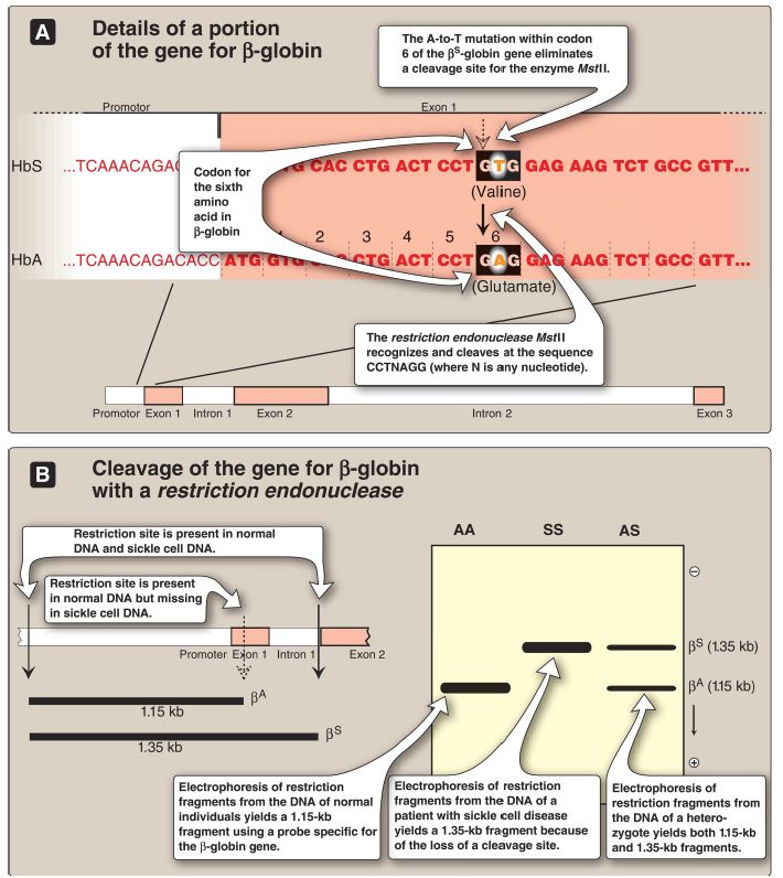
Figure 2: Detection of βS-globin mutation. kb = kilobase (1 kb = 1,000 base pairs in double-stranded DNA); Hb = hemoglobin.
a. Early diagnostic efforts: In the past, prenatal diagnosis of sickle cell anemia involved the determination of the amount and kinds of Hb synthesized in the nucleated red cells obtained from fetal blood. However, the invasive procedures to obtain fetal blood have a high mortality rate (~5%), and analysis cannot be carried out until late in the second trimester of pregnancy when HbA (and its HbS variant) begins to be produced.
b. RFLP analysis: In sickle cell anemia, the sequence alteration caused by the point mutation abolishes the recognition site of the restriction endonuclease MstII: CCTNAGG (where N is any nucleotide; see Fig. 2). Thus, the A-to-T mutation in codon 6 of the βS-globin gene eliminates a cleavage site for the enzyme. Normal DNA digested with MstII yields a 1.15-kb fragment, whereas a 1.35-kb fragment is generated from the βS gene as a result of the loss of one MstII cleavage site. Diagnostic techniques that allow analysis of fetal DNA from amniotic cells or chorionic villus sampling rather than fetal blood have proved valuable because they provide safe, early detection of sickle cell anemia as well as other genetic diseases. [Note: Genetic disorders caused by insertions or deletions between two restriction sites, rather than by the creation or loss of cleavage sites, will also display RFLP.]
4. Indirect diagnosis of phenylketonuria using RFLP:
The gene for phenylalanine hydroxylase (PAH), the enzyme deficient in phenylketonuria ([PKU]), is located on chromosome 12. It spans ~90 kb of genomic DNA and contains 13 exons separated by introns (Fig. 3). Mutations in the PAH gene usually do not directly affect any restriction endonuclease recognition site. To establish a diagnostic protocol for PKU, DNA from family members of the affected individual must be analyzed. The goal is to identify genetic markers (RFLP) that are tightly linked to the disease trait. Once these markers are identified, RFLP analysis can be used to carry out prenatal diagnosis.

Figure 3: The gene for phenylalanine hydroxylase showing 13 exons, restriction sites, and some of the >500 mutations causing phenylketonuria.
a. Mutant gene identification: Determining the presence of the mutant gene by identifying the polymorphism marker can be done if two conditions are satisfied. First, if the polymorphism is closely linked to a disease-producing mutation, the defective gene can be traced by detection of the RFLP. For example, if DNA from a family carrying a disease-causing gene is examined by restriction enzyme cleavage and Southern blotting, it is sometimes possible to find an RFLP that is consistently associated with that gene (that is, they show close linkage and are coinherited). It is then possible to trace the inheritance of the gene within a family without knowledge of the nature of the genetic defect or its precise location in the genome. [Note: The polymorphism may be known from the study of other families with the disorder or may be discovered to be unique in the family under investigation.]
Second, for autosomal-recessive disorders, such as PKU, the presence of an affected individual in the family would aid in the diagnosis. This individual would have the mutation present on both chromosomes, allowing identification of the RFLP associated with the genetic disorder.
b. RFLP analysis: The presence of abnormal genes for PAH can be shown using DNA polymorphisms as markers to distinguish between normal and mutant genes. For example, Figure 4 shows a typical pattern obtained when DNA from members of an affected family is cleaved with an appropriate restriction enzyme and subjected to electrophoresis. The vertical arrows represent the cleavage sites for the restriction enzyme used. The presence of a polymorphic site creates fragment “b” in the autoradiogram (after hybridization with a labeled PAH-cDNA probe), whereas the absence of this site yields only fragment “a.” Note that subject II-2 demonstrates that the polymorphism, as shown by the presence of fragment “b,” is associated with the mutant gene. Therefore, in this particular family, the appearance of fragment “b” corresponds to the presence of a polymorphic site that marks the abnormal gene for PAH. The absence of fragment “b” corresponds to having only the normal gene. In Figure 4, examination of fetal DNA shows that the fetus inherited two abnormal genes from the parents and, therefore, has PKU.

Figure 4: Analysis of restriction fragment length polymorphism in a family with a child affected by phenylketonuria (PKU), an autosomal-recessive disease.
The molecular defect in the gene for phenylalanine hydroxylase (PAH) in the family is not known. The family wanted to know if the current pregnancy would be affected by PKU.
c. Value of DNA testing: DNA-based testing is useful not only in determining if an unborn fetus is affected by PKU but also in detecting unaffected carriers of the mutated gene to aid in family planning. [Note: PKU is treatable by dietary restriction of phenylalanine. Early diagnosis and treatment are essential in preventing severe neurologic damage in affected individuals.]
 الاكثر قراءة في الكيمياء الحيوية
 اخر الاخبار
اخبار العتبة العباسية المقدسة

الآخبار الصحية


مواضيع ذات صلة
 قسم الشؤون الفكرية يصدر كتاباً يوثق تاريخ السدانة في العتبة العباسية المقدسة
 "المهمة".. إصدار قصصي يوثّق القصص الفائزة في مسابقة فتوى الدفاع المقدسة للقصة القصيرة
 (نوافذ).. إصدار أدبي يوثق القصص الفائزة في مسابقة الإمام العسكري (عليه السلام)


