


النبات

مواضيع عامة في علم النبات
الجذور - السيقان - الأوراق
النباتات الوعائية واللاوعائية
البذور (مغطاة البذور - عاريات البذور)
الطحالب
النباتات الطبية
الحيوان
مواضيع عامة في علم الحيوان
علم التشريح
التنوع الإحيائي
البايلوجيا الخلوية
الأحياء المجهرية
البكتيريا
الفطريات
الطفيليات
الفايروسات
علم الأمراض
الاورام
الامراض الوراثية
الامراض المناعية
الامراض المدارية
اضطرابات الدورة الدموية
مواضيع عامة في علم الامراض
الحشرات
التقانة الإحيائية
مواضيع عامة في التقانة الإحيائية
التقنية الحيوية المكروبية
التقنية الحيوية والميكروبات
الفعاليات الحيوية
وراثة الاحياء المجهرية
تصنيف الاحياء المجهرية
الاحياء المجهرية في الطبيعة
أيض الاجهاد
التقنية الحيوية والبيئة
التقنية الحيوية والطب
التقنية الحيوية والزراعة
التقنية الحيوية والصناعة
التقنية الحيوية والطاقة
البحار والطحالب الصغيرة
عزل البروتين
هندسة الجينات
التقنية الحياتية النانوية
مفاهيم التقنية الحيوية النانوية
التراكيب النانوية والمجاهر المستخدمة في رؤيتها
تصنيع وتخليق المواد النانوية
تطبيقات التقنية النانوية والحيوية النانوية
الرقائق والمتحسسات الحيوية
المصفوفات المجهرية وحاسوب الدنا
اللقاحات
البيئة والتلوث
علم الأجنة
اعضاء التكاثر وتشكل الاعراس
الاخصاب
التشطر
العصيبة وتشكل الجسيدات
تشكل اللواحق الجنينية
تكون المعيدة وظهور الطبقات الجنينية
مقدمة لعلم الاجنة
الأحياء الجزيئي
مواضيع عامة في الاحياء الجزيئي
علم وظائف الأعضاء
الغدد
مواضيع عامة في الغدد
الغدد الصم و هرموناتها
الجسم تحت السريري
الغدة النخامية
الغدة الكظرية
الغدة التناسلية
الغدة الدرقية والجار الدرقية
الغدة البنكرياسية
الغدة الصنوبرية
مواضيع عامة في علم وظائف الاعضاء
الخلية الحيوانية
الجهاز العصبي
أعضاء الحس
الجهاز العضلي
السوائل الجسمية
الجهاز الدوري والليمف
الجهاز التنفسي
الجهاز الهضمي
الجهاز البولي
المضادات الميكروبية
مواضيع عامة في المضادات الميكروبية
مضادات البكتيريا
مضادات الفطريات
مضادات الطفيليات
مضادات الفايروسات
علم الخلية
الوراثة
الأحياء العامة
المناعة
التحليلات المرضية
الكيمياء الحيوية
مواضيع متنوعة أخرى
الانزيمات

Eukaryotic DNA Organization
 المؤلف:
Denise R. Ferrier
المؤلف:
Denise R. Ferrier
 المصدر:
Lippincott Illustrated Reviews: Biochemistry
المصدر:
Lippincott Illustrated Reviews: Biochemistry
 الجزء والصفحة:
 22-12-2021
 2124
2124


+
-
20
Eukaryotic DNA Organization
A typical (diploid) human somatic cell contains 46 chromosomes, whose total DNA is ~2 m long! It is difficult to imagine how such a large amount of genetic material can be effectively packaged into a volume the size of a cell nucleus so that it can be efficiently replicated and its genetic information expressed. To do so requires the interaction of DNA with a large number of proteins, each of which performs a specific function in the ordered packaging of these long molecules of DNA. Eukaryotic DNA is associated with tightly bound basic proteins, called histones. These serve to order the DNA into fundamental structural units, called nucleosomes, which resemble beads on a string.
Nucleosomes are further arranged into increasingly more complex structures that organize and condense the long DNA molecules into chromosomes that can be segregated during cell division. [Note: The complex of DNA and protein found inside the nuclei of eukaryotic cells is called chromatin.]
A. Histones and nucleosome formation
There are five classes of histones, designated H1, H2A, H2B, H3, and H4. These small, evolutionally conserved proteins are positively charged at physiologic pH as a result of their high content of lysine and arginine. Because of their positive charge, they form ionic bonds with negatively charged DNA. Histones, along with ions such as Mg2+, help neutralize the negatively charged DNA phosphate groups.
1. Nucleosomes: Two molecules each of H2A, H2B, H3, and H4 form the octameric core of the individual nucleosome “beads.” Around this structural core, a segment of dsDNA is wound nearly twice (Fig. 1).
Winding eliminates a helical turn, causing negative supercoiling. [Note: The N-terminal ends of these histones can be acetylated, methylated, or phosphorylated. These reversible covalent modifications influence how tightly the histones bind to the DNA, thereby affecting the expression of specific genes. Histone modification is an example of epigenetics, or heritable changes in gene expression caused without alteration of the nucleotide sequence.] Neighboring nucleosomes are joined by linker DNA ~50 bp long. H1 is not found in the nucleosome core, but instead binds to the linker DNA chain between the nucleosome beads. H1 is the most tissue specific and species specific of the histones. It facilitates the packing of nucleosomes into more compact structures.

Figure 1: Organization of human DNA, illustrating the structure of nucleosomes. H = histone.
2. Higher levels of organization: Nucleosomes can be packed more tightly (stacked) to form a nucleofilament. This structure assumes the shape of a coil, often referred to as a 30-nm fiber. The fiber is organized into loops that are anchored by a nuclear scaffold containing several proteins. Additional levels of organization lead to the final chromosomal structure (Fig. 2).
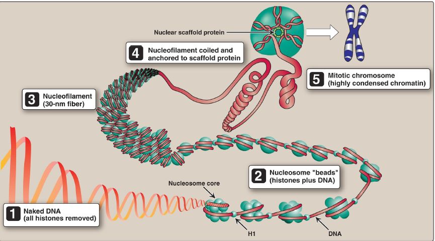
Figure 2 Structural organization of eukaryotic DNA. [Note: A 104 linear compaction is seen from 1–5.] H = histone.
B. Nucleosome fate during DNA replication
Parental nucleosomes are disassembled to allow access to DNA during replication. Once DNA is synthesized, nucleosomes form rapidly. Their histone proteins come both from de novo synthesis and from the transfer of parental histones.
 الاكثر قراءة في الكيمياء الحيوية
 اخر الاخبار
اخبار العتبة العباسية المقدسة

الآخبار الصحية


مواضيع ذات صلة
 قسم الشؤون الفكرية يصدر كتاباً يوثق تاريخ السدانة في العتبة العباسية المقدسة
 "المهمة".. إصدار قصصي يوثّق القصص الفائزة في مسابقة فتوى الدفاع المقدسة للقصة القصيرة
 (نوافذ).. إصدار أدبي يوثق القصص الفائزة في مسابقة الإمام العسكري (عليه السلام)


