


النبات

مواضيع عامة في علم النبات
الجذور - السيقان - الأوراق
النباتات الوعائية واللاوعائية
البذور (مغطاة البذور - عاريات البذور)
الطحالب
النباتات الطبية
الحيوان
مواضيع عامة في علم الحيوان
علم التشريح
التنوع الإحيائي
البايلوجيا الخلوية
الأحياء المجهرية
البكتيريا
الفطريات
الطفيليات
الفايروسات
علم الأمراض
الاورام
الامراض الوراثية
الامراض المناعية
الامراض المدارية
اضطرابات الدورة الدموية
مواضيع عامة في علم الامراض
الحشرات
التقانة الإحيائية
مواضيع عامة في التقانة الإحيائية
التقنية الحيوية المكروبية
التقنية الحيوية والميكروبات
الفعاليات الحيوية
وراثة الاحياء المجهرية
تصنيف الاحياء المجهرية
الاحياء المجهرية في الطبيعة
أيض الاجهاد
التقنية الحيوية والبيئة
التقنية الحيوية والطب
التقنية الحيوية والزراعة
التقنية الحيوية والصناعة
التقنية الحيوية والطاقة
البحار والطحالب الصغيرة
عزل البروتين
هندسة الجينات
التقنية الحياتية النانوية
مفاهيم التقنية الحيوية النانوية
التراكيب النانوية والمجاهر المستخدمة في رؤيتها
تصنيع وتخليق المواد النانوية
تطبيقات التقنية النانوية والحيوية النانوية
الرقائق والمتحسسات الحيوية
المصفوفات المجهرية وحاسوب الدنا
اللقاحات
البيئة والتلوث
علم الأجنة
اعضاء التكاثر وتشكل الاعراس
الاخصاب
التشطر
العصيبة وتشكل الجسيدات
تشكل اللواحق الجنينية
تكون المعيدة وظهور الطبقات الجنينية
مقدمة لعلم الاجنة
الأحياء الجزيئي
مواضيع عامة في الاحياء الجزيئي
علم وظائف الأعضاء
الغدد
مواضيع عامة في الغدد
الغدد الصم و هرموناتها
الجسم تحت السريري
الغدة النخامية
الغدة الكظرية
الغدة التناسلية
الغدة الدرقية والجار الدرقية
الغدة البنكرياسية
الغدة الصنوبرية
مواضيع عامة في علم وظائف الاعضاء
الخلية الحيوانية
الجهاز العصبي
أعضاء الحس
الجهاز العضلي
السوائل الجسمية
الجهاز الدوري والليمف
الجهاز التنفسي
الجهاز الهضمي
الجهاز البولي
المضادات الميكروبية
مواضيع عامة في المضادات الميكروبية
مضادات البكتيريا
مضادات الفطريات
مضادات الطفيليات
مضادات الفايروسات
علم الخلية
الوراثة
الأحياء العامة
المناعة
التحليلات المرضية
الكيمياء الحيوية
مواضيع متنوعة أخرى
الانزيمات

The Cloning Process
 المؤلف:
John M Walker and Ralph Rapley
المؤلف:
John M Walker and Ralph Rapley
 المصدر:
Molecular Biology and Biotechnology 5th Edition
المصدر:
Molecular Biology and Biotechnology 5th Edition
 الجزء والصفحة:
 17-11-2020
 3192
3192


+
-
20
The Cloning Process
The recombinant vector molecule must be introduced into its ‘matching’ host cell in order to replicate and produce multiple copies. The process by which DNA is introduced into the host cell is known as bacterial transformation. Since ‘naked’ vector DNA is hydrophilic and the bacterial cell wall is normally impermeable to such molecules, the host cell must be made ‘competent’ by treatment with calcium chloride in the early log phase of growth. This causes the cell to become permeable to chloride ions. When competent cells are mixed with DNA and heat shocked at 42 °C, the swollen cells are able to take up the naked DNA molecules. It is believed that only a single DNA molecule is permitted to enter any single cell. Thus individual colonies of transformed bacteria grow single recombinant vector molecules. The bacterial cells are usually grown on selective media so that only transformants survive to form colonies. Thus, for example, if the vector contains an ampicillin resistance gene and the cells are grown on ampicillin-containing media, only the cells containing the vector will form colonies.
1- Library Screening
There are several methods of screening for transformed colonies that contain recombinant vectors. For example, where the cloning site in the vector lies within an antibiotic resistance gene, successful integration of the insert will lead to inactivation of the resistance gene and recombinant colonies can be identified by a technique known as replica plating (Figure 1). In this method, the pattern of colonies in the original Petri dish is printed on to a nutrient agar plate containing the selective antibiotic.
The position of the recombinant colonies, i.e. those that fail to grow on the selective antibiotic, is noted so that they can be picked from the master plate. In other vectors, a method called blue/white selection can be performed. In blue/white selection, successful integration of a foreign DNA molecule in the vector destroys an enzyme gene (the LacZ gene of b-galactosidase) that otherwise forms a blue product when the transformed colonies are exposed to the substrate X-gal. Thus recombinant colonies are white and non-recombinants are blue (Figure 2).
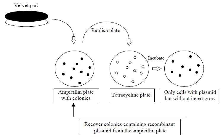
Figure 1: Replica plating to detect recombinant plasmids. A sterile velvet pad is pressed on to the surface of an agar plate, picking up some cells from each colony growing on that plate. The pad is then pressed on to a fresh agar plate containing the selective antibiotic, thus inoculating it with cells in a pattern identical with that of the original colonies. Clones of cells that fail to grow on the second plate (owing to the loss of antibiotic resistance) can be recovered from their corresponding colonies on the first plate.
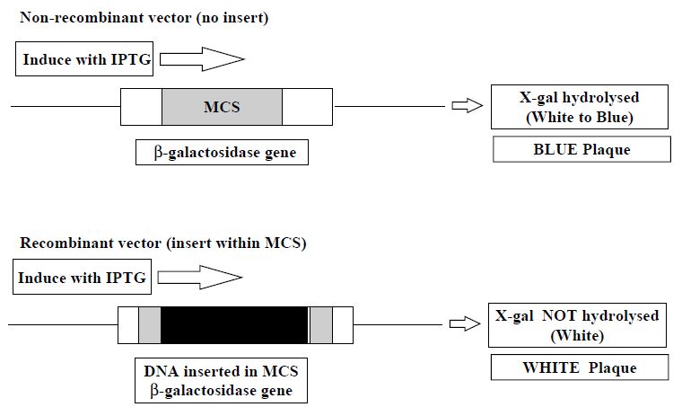
Figure 2: Principle of blue/white selection for the detection of recombinant vectors. In the presence of the inducer IPTG the b-galactosidase (LacZ) gene is transcribed. The recombinant DNA insert disrupts the expression of the LacZ gene which encompasses the multiple cloning site (MCS), hence the substrate X-gal is not hydrolysed and the recombinant colonies remain white.
2- Identifying Clones
Bacterial clones containing the sought after recombinant vectors can be identified by hybridisation with specific radioactively labelled or enzyme-labelled cDNA or genomic DNA probes or alternatively by the immunodetection of protein products (using specialised expression vectors which allow a cloned foreign cDNA to be transcribed to express its protein product). Both approaches are technically straightforward.
Both involve the transfer of bacterial colonies from a master agar plate on to carefully orientated nitrocellulose or nylon membranes. The cells are then lysed and the DNA (or protein) from the lysed colonies is immobilised on the membrane, which is used for the probing step.
Recombinant colonies can be detected as spots on X-ray film either by autoradiography or enzyme-generated chemiluminescence. The spots on the X-ray film can then be aligned with the agar master plate allowing the correct colonies to be picked (Figure 5.5). For protein detection in expression vectors, antibody probes are employed and in a manner analogous to an ELISA test. The antibody probe is conjugated to an enzyme such as horseradish peroxidase or alkaline phosphatase. It is the activity of the bound enzyme on its chemiluminescence or chromogenic substrate that reveals the position of the recombinant colonies.
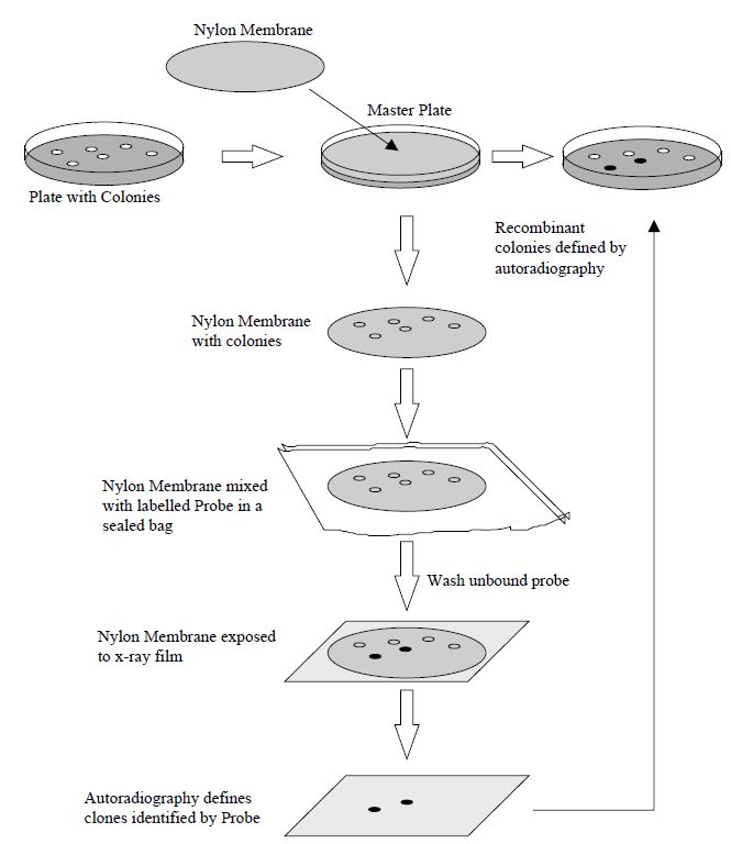
Figure 3: Method for detection of recombinant clones by colony hybridisation with labelled gene probes. The bacteria immobilised on the nylon (or nitrocellulose) membrane are lysed in alkaline conditions to make the plasmid DNA accessible to the probe.
 الاكثر قراءة في مواضيع عامة في الاحياء الجزيئي
 اخر الاخبار
اخبار العتبة العباسية المقدسة

الآخبار الصحية


مواضيع ذات صلة
 قسم الشؤون الفكرية يصدر كتاباً يوثق تاريخ السدانة في العتبة العباسية المقدسة
 "المهمة".. إصدار قصصي يوثّق القصص الفائزة في مسابقة فتوى الدفاع المقدسة للقصة القصيرة
 (نوافذ).. إصدار أدبي يوثق القصص الفائزة في مسابقة الإمام العسكري (عليه السلام)


